Archive : Article / Volume 2, Issue 1
- Research Article | DOI:
- https://doi.org/10.58489/2837-3367/003
A novel technique of ear and nose piercing by intravenous cannula
- Assistant professor, ENT and HNS, Khulna City Medical college, Khulna.
- Professor and Head of Department, ENT and HNS, Khulna City Medical college, Khulna.
- Resident, Anaesthesiology and ICU, Jashore medical college, Jashore.
- Assistant professor. ENT and HNS, Prime Medical college, Rangpur.
Saikat Bhattacharjee
Saikat Bhattacharjee, Sudhangshu K.B, Prianka C, Monwar M.B., (2023). A novel technique of ear and nose piercing by intravenous cannula. Journal of ENT and Healthcare. 2(1). DOI: 10.58489/2837-3367/003
© 2023 Saikat Bhattacharjee, this is an open access article distributed under the Creative Commons Attribution License, which permits unrestricted use, distribution, and reproduction in any medium, provided the original work is properly cited.
- Received Date: 07-01-2023
- Accepted Date: 22-12-2022
- Published Date: 10-01-2023
Ear, nose, piercing, intravenous cannula, technique
Abstract
Background:
Ear and nose piercing has been a common practice in almost all societies around the globe since beginning of human civilization. It is done to wear ornaments for beautification. Beauticians are practicing the procedure by sewing needle and piercing gun that causes various complications. Newer techniques are emerging to minimise complications. We have described a new technique of ear and nose piercing which minimized complications.
Objective:
Our aim is to describe the new technique and to study the outcome of the procedure.
Method:
A total 32 cases who have undergone ear and or nose piercing were included in this study. All cases were selected from ENT outpatient department of a medical college hospital and private consultation clinic during July 2021 to June 2022.
Results:
In all cases ear and or nose piercing were done by our new technique by using intravenous cannula. Cases were followed up to 6 months periodically and found no immediate or late complications.
Conclusion:
Ear and nose piercing by intravenous cannula is simple, cost effective and well accepted by the patients.
Introduction
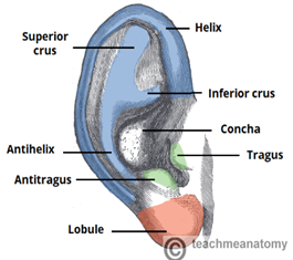
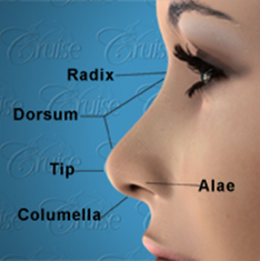
Ear and nose piercing is commonly practiced in our subcontinent and many other countries for wearing ornaments mostly by female and uncommonly by male [3]. It is also very common among tribal people for their own social and cultural tradition. In Bangladesh ear and nose piercing is common in almost all female and least common in male. Traditional tools like sewing needle and thorn are used for the procedure [2]. Now-a-days piercing gun and piercing kits are available and used by beauticians [4]. The pinna of external ear is made up of single cartilage covered by skin. The auricle has tragus, antitragus, helix, antihelix, concha, and lobule [6]. The lobule is devoid of cartilage, composed of loose areolar tissue, fat and is the preferred site for piercing. External nose is also composed of cartilage and bone. The alae of external nose and columella of septum are the sites for piercing. Reported complications of ear and nose piercing are wound infection, granuloma formation, keloid, and split ear lobule. Complications mostly happened when traditional methods are used.


Objective
Our aim is to assess the outcome of ear and nose piercing by intravenous cannula instead of sewing needle, thorn, piercing gun.
Method
This study enrolled 32 cases during the period of two year starting from July 2020 to June 2022. All cases were selected from ENT outpatient department Khulna City Medical College Hospital and from private consultation of the authors.
Inclusion criteria was female patients with no previous history of ear and or nose piercing with complications of infection, granuloma, or keloid formation.
Exclusion criteria was male patient, female patient with previous history of ear and or nose piercing with complications and patients who asked for piecing in cartilaginous part of pinna.
The patients were selected for outdoor procedure without any anaesthesia. We have used 14G or 16G intravenous cannula for piercing. With all aseptic precautions after inserting the cannula at selected point on ear lobule and or septal columella, alae of external nose, the trocar is withdrawn [3,4]. Then the cannula is cut keeping approximately 1 centimeter at both outside ends. Povidone 5% topical ointment was advised to apply around the cannula thrice daily [8]. On fourth day after removal of the canula gold made ear or nose ring was wearied and clasp of the stud fitted properly avoiding any undue pressure on pierced site. Topical povidone iodine ointment continued for 2 weeks. Patient was advised to rotate the stud of the ear or nose ring after applying povidone iodine ointment. All cases were followed up to 6 months periodically for any complications.
Results
All cases included in this study were female age ranging from 8years to 30years with a mean age of 19yearsAll cases came from town dwellers, students and from good socioeconomic status with good educational background. Not a single patient developed any complications like wound infection, keloid formation, ear lobule or columellar split. Two patients (6.25%) who wearing gold looking imitation ear ring developed foreign body granuloma that was managed by replacing the imitation ring by gold ring and applying antibiotic-steroid ointment. In two cases (6.25%) the cannula was expelled out accidentally on second day. We reinserted the cannula, negotiated proline thread through the cannula and secured by knot.
Discussion
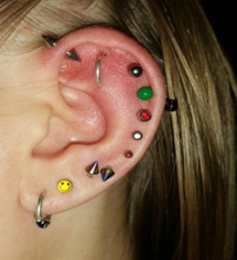
The practice of body piercing particularly ear and nose piecing is increasing among the female. This is also gaining popularity among young male too imitating the celebrities. In addition to piercing in ear lobule transcartilaginous ear piercing has been gaining popularity.

In our experience we have observed an increase the incidence of auricular perichondritis, perichondrial abscess followed by auricular deformity and keloid formation after trans cartilaginous piercing [8,9].
There are various techniques of piercing. There are different methods of ear and nose piercing. they are sewing needle technique, intravenous canula stent, spring loaded piercing gun and many more [5]. All of them have disadvantage. Piercing gun is associated with embedded earrings [6,7]. The main targets of any surgical technique are that it should be easy, reproducible, cost effective, accurate and should create an epithelialized straight hole without any complications [1]. Our technique is simple and reproducible for ear and nose piercing by an easily available, sterile intravenous cannula. The stud of the earring can easily be negotiated through the cannula after removing the needle.
Traditional method is by ordinary sewing needle and thread, surgical proline suture needle. Recently piecing gun and piercing kits are available which are being used by beauticians in the parlour. Majority cases develop wound infection, granuloma, hypertrophied scar and keloid. Post procedure care is also an important aspect. The earrings should be kept in place until the tract has epithelialized which requires 2 to 3 weeks. Long term use of heavy jewellery may cause elongation of the hole leading to ear lobule split.
Our technique of piercing by intravenous cannula is the modification of traditional needle technique and is free from complications.
Conclusion
In our study ear and nose piercing by intravenous canula technique is found to be simpler, cost effective and free from complications than other traditional methods and piercing gun. It is also satisfactory after repair of split ear lobule. Trancartilaginous piercing is discouraged. Follow up revealed no complications. So, we do recommend this procedure.
Conflict of interest
none
Sponsor
None.
References
- Saple, A. (2022), Ear Piercing—a Simple Solution. Indian J Surg 84, 242–244.
- Sirunya Silapunt MD, Leonard H Goldberg MD - Surgery of the Skin - 2005, Chapter 20 - Repair of the Split Earlobe, Ear Piercing, and Earlobe Reduction, Pages 291-309
- Tiwari VK, Misra A, Sarabahi S. A novel technique for piercing of ear lobule suited to Indian subcontinent. Indian J Plast Surg. 2013; 46:160–1.
- Landeck A, Newman N, Breadon J, Zahner S. A simple technique for ear piercing. Journal of the American Academy of Dermatology.
- Zackowski DA. An IV cannula stent for ear piercing. Plast Reconstr Surg. 1987; 80 ([letter]): 751
- Cockin J, Finan P, Powell M. A problem with ear piercing. Br Med J. 1977; 2: 1631
- Saleeby ER, Rubin MG, Youshock E, Kleinsmith DM. Embedded foreign bodies presenting as earlobe keloids. J Dermatol Surg Oncol. 1984; 10: 902-904
- Widick MH, Coleman J. Perichondrial abscess resulting from a high ear-piercing: case report. Otolaryngol Head Neck Surg. 1992; 107: 803-804
- Hendricks WM. Complications of ear piercing: treatment and prevention. Cutis. 1991; 48: 386-394
- Watson MG, Campbell J B, Pahor A L. Complications of nose piercing. Br Med J. 1987 May 16; 294(6582): 1262.


