Archive : Article / Volume 2, Issue 1
- Case Report | DOI:
- https://doi.org/10.58489/2836-5127/004
A Subdural Supratentorial Hemorrhage and Contusion after a Traumatic Brain Injury Mimicking a Dural Thickening: A Clinical Case
Radiology Specialist, Department of Radiology, Al-Namas General Hospital, Ministry of Health, Al-Namas City, Saudi Arabia.
Abdulwahab Alahmari
Abdulwahab Alahmari, (2022). A Subdural Supratentorial Hemorrhage and Contusion after a Traumatic Brain Injury Mimicking a Dural Thickening: A Clinical Case. Journal of Radiology Research and Diagnostic Imaging .2(1). DOI: 10.58489/2836-5127/004.
© 2022 Abdulwahab Alahmari, this is an open access article distributed under the Creative Commons Attribution License, which permits unrestricted use, distribution, and reproduction in any medium, provided the original work is properly cited.
- Received Date: 20-12-2022
- Accepted Date: 05-01-2023
- Published Date: 10-01-2023
Abstract
This is a paper about a case of a subdural hemorrhage and contusion mimicking a dural thickening. The bone window was applied which helped in showing the bleeding very clear. This case will discuss this case and the usefulness of the bone window in similar scenarios.
Case Report
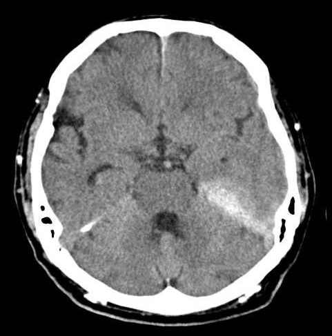
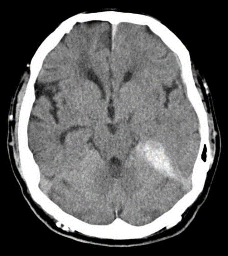
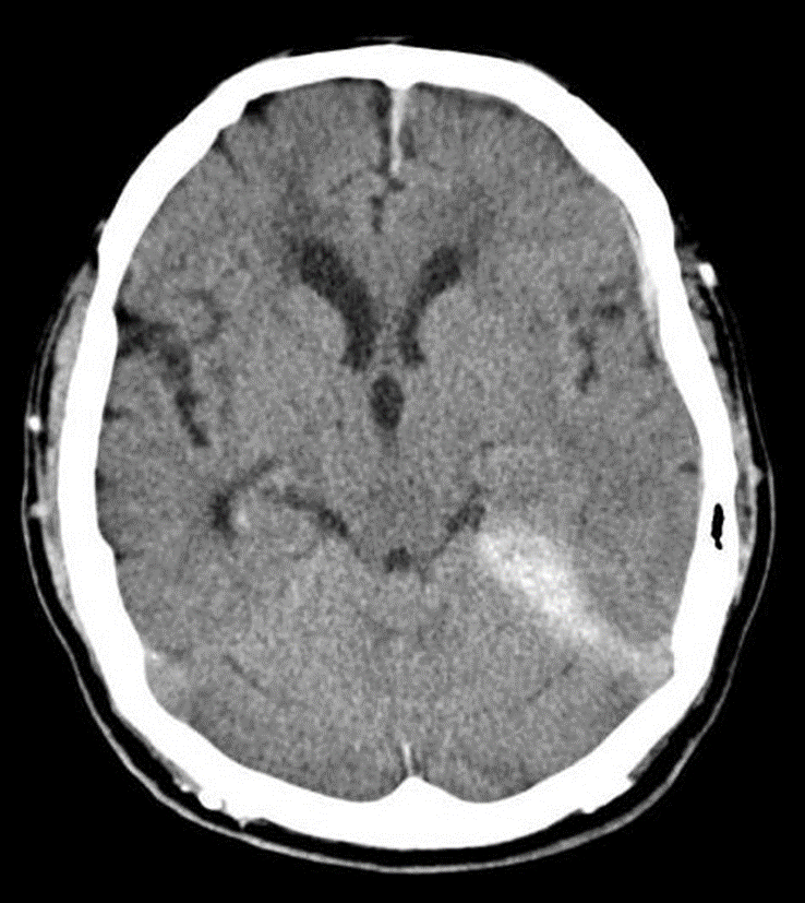
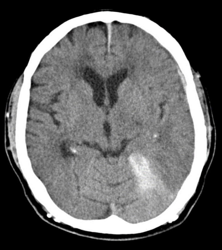
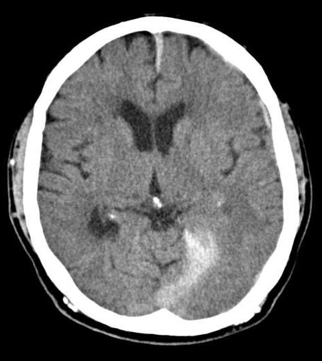
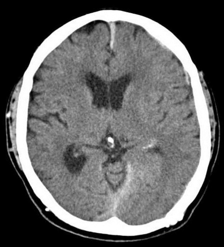
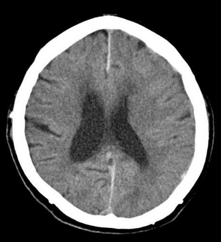
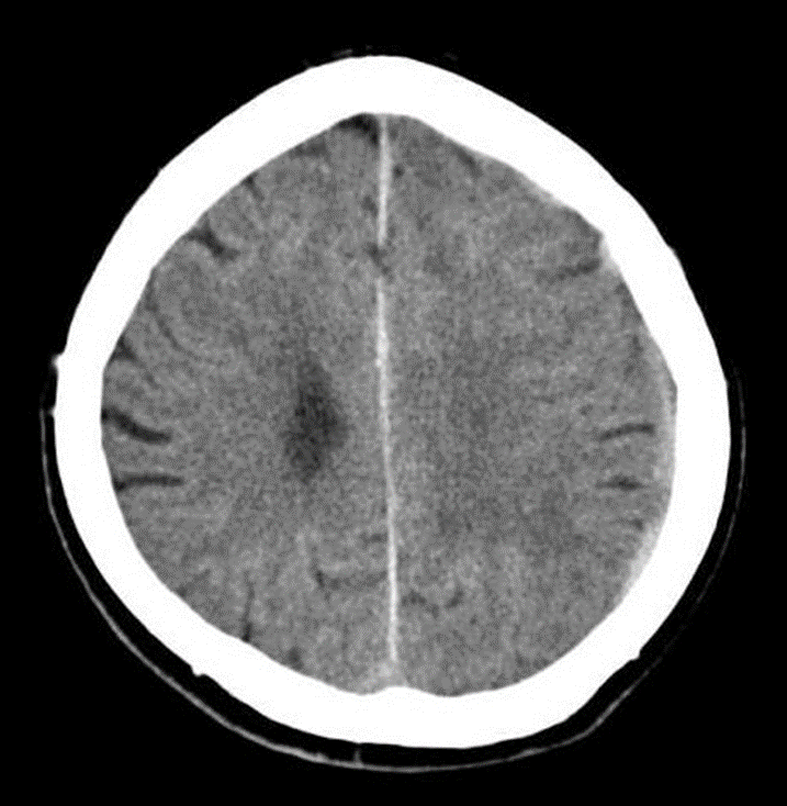
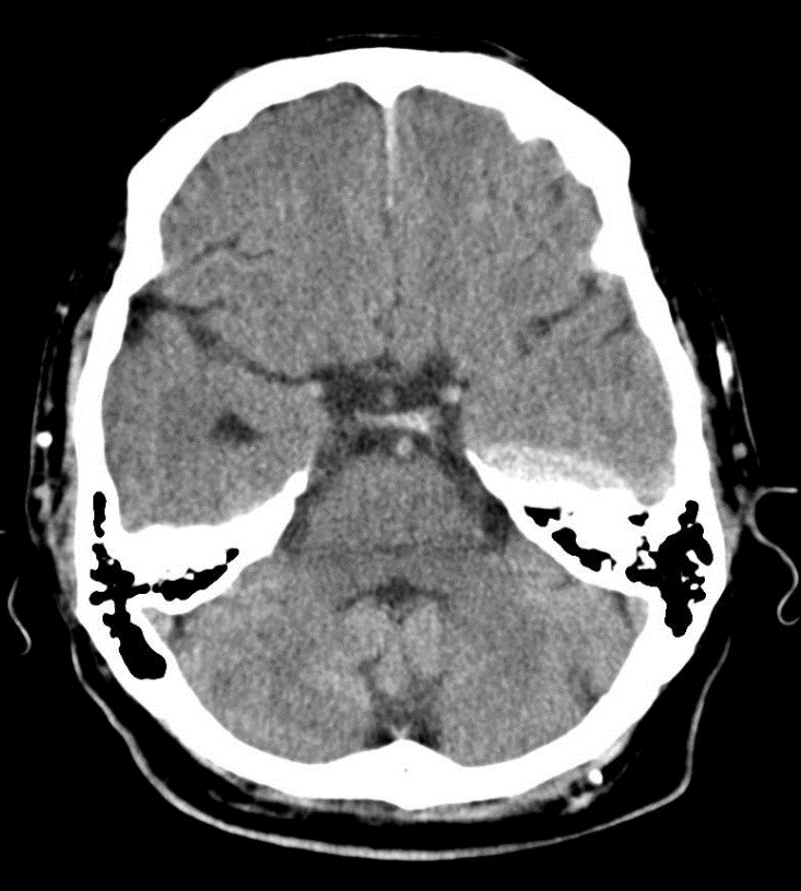
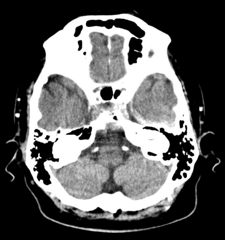
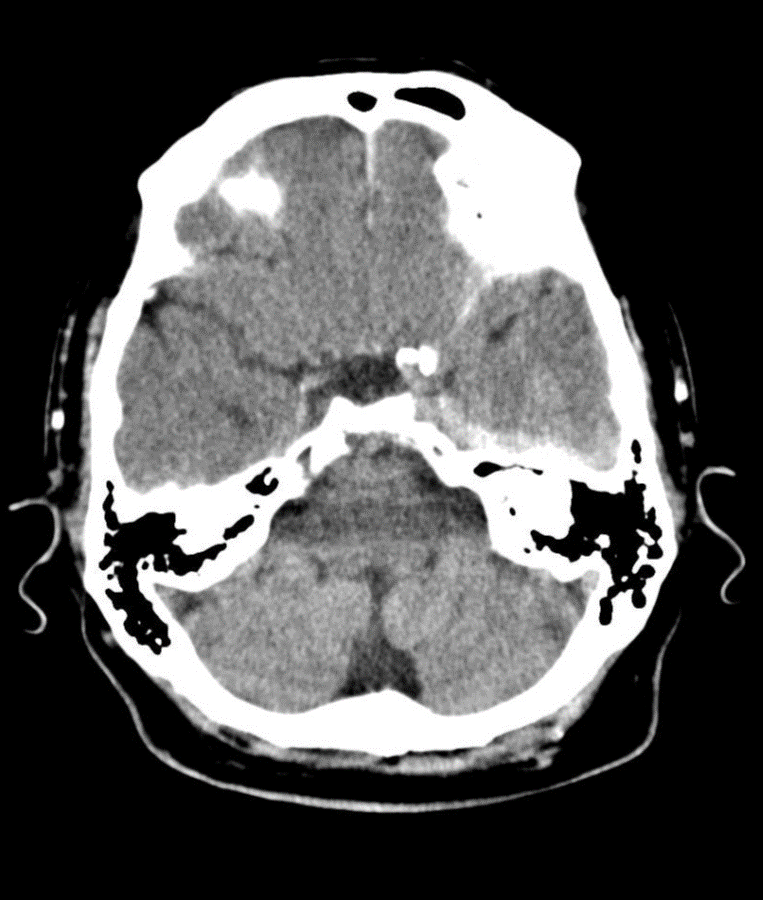
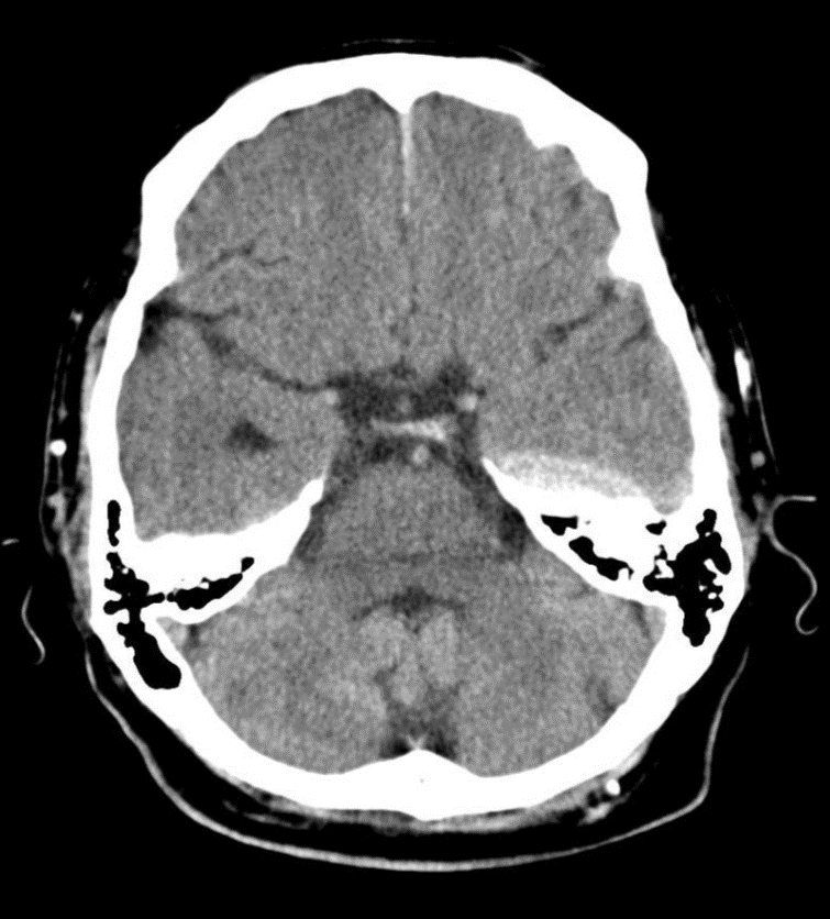
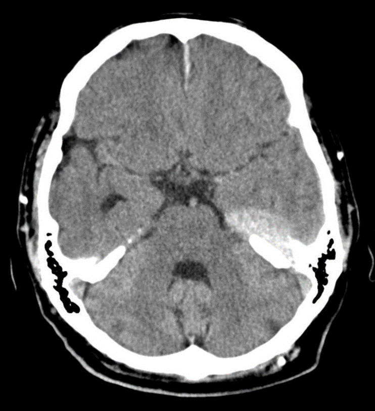
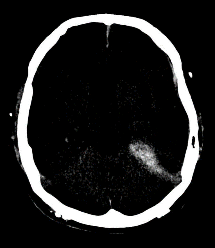
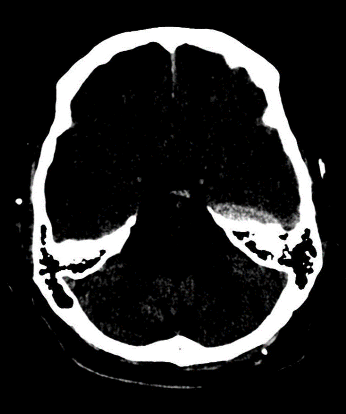
A 46-year-old male patient came to the emergency room after a falling down on his back from a short distance and his head hit the ground. The patient felt severe headache and came to the emergency room. The patient used to take anticoagulant medications and fall down on his head. A CT scan of the brain was requested to check the patient’s brain. The CT scan showed a diffuse white mass extended from below the left temporal lobe to above the left tentorium. The white dense area looks like a dural thickening. By adjusting the CT window’s contrast similar to the region of interest (ROI), the white dense area looks like a continuation of one mass extending from the temporal bone to the tentorium. The brain CT revealed a a supra-tentorium cerebelli, falx cereberi, and temporal lobe subdural hemorrhage and contusion on the left side see (Figs. from 1 thru 15).
Discussion
Supratentorial subdural hemorrhage is a rare finding [1]. Causes of such cases are: tentorial tear, brain contusion, extension of interhemispheric hemorrhage, and bridging veins injuries [1]. Deformation of the cranium during delivery can causes tentorial tearing, but in this case of an adult who never had any issues at birth is unlikely scenario. Pial and dural bridging veins penetrate the tentorial form the lateral side [1]. Tentorial hemorrhage was noticed in newborns who undergo a vacuum extraction and those newborns experienced seizures [2]. In a case of aneurysm of the anterior communicated artery which caused a symmetrical tentorial thickening of dura mater bilaterally [3]. The prognosis of supratentorial hemorrhage cases when surgical intervention is used, it depends mainly on the size of the hemorrhage. As more the size of the hemorrhage increases, as the prognosis becomes worse [4].
Conclusion
Using of bone window can help in showing brain hemorrhage. Usually, supratentorial hemorrhage is common in newborns and children.
References
- MATSUMOTO K, HOURI T, YAMAKI T, UEDA S. (1996). Traumatic Acute Subdural Hematoma Localized on the Superior Surface of the Tentorium Cerebelli—Two Case Reports—. Neurologia medico-chirurgica.;36(6):377-9.
- Huang, C. C., & Shen, E. Y. (1991). Tentorial subdural hemorrhage in term newborns: ultrasonographic diagnosis and clinical correlates. Pediatric neurology, 7(3), 171-177.
- Krishnaney, A. A., Rasmussen, P. A., & Masaryk, T. (2004). Bilateral tentorial subdural hematoma without subarachnoid hemorrhage secondary to anterior communicating artery aneurysm rupture: a case report and review of the literature. American journal of neuroradiology, 25(6), 1006-1007.
- Rehman, W. A., & Anwar, M. S. (2017). Surgical outcome of spontaneous supra tentorial intracerebral hemorrhage. Pakistan journal of medical sciences, 33(4), 804.


