Archive : Article / Volume 1, Issue 2
- Research Article | DOI:
- https://doi.org/10.58489/2836-2179/007
Age Characteristics of Fatigue During Cyclic Work of Maximum Power
Vasil Stefanic National University, Department of physical education and Sport, Ukraine.
Serhii Popel
Serhii Popel, Roman Faychak, irina Tcap, Ivano-Frankivsk, (2022). Age Characteristics of Fatigue During Cyclic Work of Maximum Power. Journal of Emergency and Nursing Management. 1(2). DOI: 10.58489/2836-2179/007
© 2022, Serhii Popel, this is an open access article distributed under the Creative Commons Attribution License, which permits unrestricted use, distribution, and reproduction in any medium, provided the original work is properly cited.
- Received Date: 30-11-2022
- Accepted Date: 16-12-2022
- Published Date: 27-12-2022
electromyography, physical activity, fatigue, age characteristics.
Abstract
The purpose of the article is a study the age-related features of changes in the functional state of the neuromuscular apparatus during cyclic work before failure in laboratory conditions. Methods:14 adult ski racers (25-28 years old) candidates for masters of sports and 12 teenagers (14-15 years old) I and II sports categories took part in the study. As the maximum physical load (before failure), an imitation of alternating two-step walking in place was used.For each subject, the walking pace was 75% of the maximum.Ski racers performed imitation under an electronic metronome. The length of the step remained unchanged throughout the study; the duration of the simulation reached 30-40 minutes.The following physiological indicators were used to assess the state of the neuromuscular apparatus: reflex excitability of spinal motoneurons (according to the amplitude of the maximum H-response); latent period of H- and M-responses; the speed of propagation of excitation along the sensory and motor fibers of the tibial nerve in the area of the popliteal fossa and the medial condyle (bone) of the tibia using the "Micro-Neuro-Soft" electroneuromyography.Results. It was established that the relative share of motoneurons participating in the reflex response is the same in resting adults and young skiers. The duration of physical exercise in teenagers reached approximately the same values ââas adult athletes and is 30-40 minutes. However, the dynamics of the studied functional indicators had their own specific features: in young ski racers, the amplitude of the H-response when refusing to continue working decreased by only 24.0% compared to the initial level. Conclusion. Reflex excitability of spinal motoneurons after performing cyclic work of maximum power in adult athletes is more pronounced than in adolescent athletes, which indicates faster fatigue after testing, but high physical performance during testing.
Introduction
Statement of the problem and analysis of the results of recent research. Fatigue as a physiological phenomenon always leads to a decrease in the efficiency of functional systems or the body as a whole (Vlasova, 2017; Hockey, 2010). Despite numerous studies (Urbanek, 2016,; Dan'ko, 2009; Kotlo, 2017; Koc, 2017) aimed at solving the problems of fatigue, questions about the factors that limit the physical capacity of people of different ages for one or another motor activity are still debatable. The main causes of fatigue during long-term physical exercises of high and maximum power are factors associated with a decrease in the level of energy supply of working muscles, as well as a violation of electrochemical reactions in working muscles and deterioration of the activity of the central nervous system in conditions of severe hyperthermia, dehydration, and metabolic disorders body balance [3,9,10]. All this indicates the complex nature of the development of fatigue. Skiing is characterized by pronounced energy expenditure, the depletion of which can cause fatigue. It is manifested by certain changes in the neuromuscular apparatus, the state of which can be juRGed by EMG indicators (Chuhlanceva, 2011; Enck, 2006; Hockey, 2010;). However, such studies in skiers were carried out sporadically and are of a fragmentary nature, which requires their updating with a review of the modern interpretation of EMG indicators.
The purpose of the research is to study the age-specific changes in the functional state of the neuromuscular apparatus during maximal cyclic work in laboratory conditions.
Material & methods. 14 adult ski racers (25-28 years old) candidates for master of sports (research group RG-1) and 12 teenagers (15-17 years old) I and II sports categories (RG-2) took part in the study. As the maximum physical load (before failure), an imitation of alternating two-step walking in place was used. For each subject, the walking pace was 75 percentage of the maximum. The imitation was performed under an electronic metronome. The length of the step remained unchanged throughout the study; the duration of the simulation reached 30-40 minutes.
The following physiological indicators were used to assess the state of the neuromuscular apparatus: reflex excitability of spinal motoneurons (according to the amplitude of the maximum H-response); latent period (LP) of H- and M-responses; speed of propagation of excitation (SPЕ) along sensitive and motor fibers of the tibial nerve (n. tibialis) in the region of the popliteal fossa and medial condyle (bone) of the tibia. This nerve provides the function of the calf muscle (m. gastrocnemius). The choice of this muscle is dictated by its exclusive participation in pushing the foot away from the support when walking and running, which is very important for skiers. Registration of the named physiological parameters was carried out according to the generally accepted method using the electroneuromyograph "Micro-Neuro-Soft" [3,12]. SPЕ on motor nerve fibers was determined by the difference in the latent periods of the M-response upon irritation of the proximal and distal points of the tibial nerve (Enck, 2006). The calculation of the SPE on sensitive nerve fibers was carried out according to the formula:

де V – speed; S – the distance between nerve irritation points; T1 – LP H-responses to stimulation of the distal point of the tibial nerve; T2 – LP H-responses when the proximal point of this nerve is irritated. The reflex response from the calf muscle was derived from monopolar surface electrodes. All studied indicators were registered before the start of the work and immediately after its completion. The amplitude of H- and M-responses was also recorded 5 minutes after the start of work.
Mathematical and statistical processing of research results was carried out in automatic mode using computer software packages that are part of the "Micro-Neuro-Soft" complex.
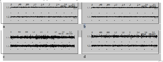
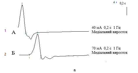
Results. In the state of muscle rest, the amplitude of the maximum H- and M-responses in skiers RG-1 and RG-2 have no probable difference (Fig. 1 a,b; Table 1). At the same time, the amplitude of the M-response has significantly larger values than in untrained people and indicates the large size of motor units [7]. Indicators of the latent period of H- and M-response in DH-1 are slightly higher compared to skiers of DH-2, which indicates a higher speed of conduction of excitation along motor fibers in adolescent athletes (Fig. 2 a) and according to the data of individual authors [3], corresponds to age characteristics.

Figure 1. Interference EMG of the right (1.1 k.) and left (1.2 k.) m.gastrocnemius, nn. tibialis, L4-5–S2 at rest (a, b) and after a one-time maximum physical load until failure: a,b – RG-1;c, d - RG-2.
Table 1. Changes in the main parameters of electroneuromyography in skiers of different ages under the influence of physical load, (M ± m, n = 26)
State | Contingent | Indicators | |||
Amplitude, mV | LP, Ms | ||||
Н-response | M-response | Н-response | M-response | ||
In peace | Adults Teenagers | 5,6±0,42 | 14,9±1,81 | 31,2±0,36 | 5,6±0,20 |
Teenagers | 5,5±0,53 | 14,7±2,53 | 29,9±0,33 | 5,2±0,18 | |
After exercise | Adults | 14,2±0,36* | 44,1±1,97* | 30,1±0,25 | 5,4±0,14 |
Teenagers | 7,3±0,41 | 21,2±2,84* | 29,4±0,22 | 4,9±0,08 | |
Note. * - the difference is probable at p<0>.
At rest, the Н/M ratio for the calf muscle in ski racers is 37.5percentage, in teenagers it is 34.7 percentage. This fact may indicate that the relative share of motoneurons participating in the reflex response without stimulation of muscle fibers is almost the same in both DH-1 and RG-2 skiers. This corresponds to the data of A.N. Huseva [3], which indicates the relative leveling of the age difference in the motor innervation of skeletal muscles after the end of the pubertal period of ontogenesis, which accelerates during intense physical exertion, especially in young athletes.


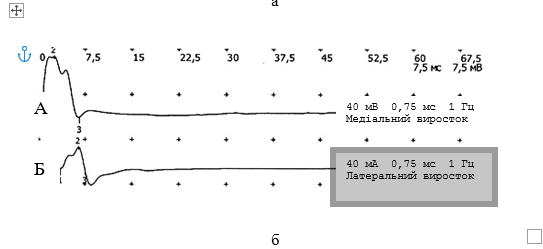
Fig. 2. Latency period and speed of propagation of excitation in m. gastrocnemius, n. tibialis, L4-5–S2, by motor (a) and sensory (b) nerve fibers in adolescent skiers (A) and adult skiers (B).
In the course of performing cyclical work, regular changes in the state of the neuromuscular apparatus were observed. Thus, the amplitude of H- and M-responses in DH-1 athletes increased 2.5 times from the initial values (p<0 xss=removed>.
After the end of the second series of the study, it was established that the duration of physical exercise in RG-2 skiers reached approximately the same values as DH-1 athletes and is 30-40 minutes. Immediately after the refusal to perform physical work, the amplitude of the maximum H-response in skiers in two groups decreased. However, relative to the initial indicators, it was still higher: in DH-1 by 23.5percentage, while in RG-2 only by 9.4 percentage (p<0 xss=removed>.
This indicates the rapid development of fatigue immediately after performing physical work and is a reflection of a general biological law: the greater the external load and force of muscle contraction, the greater the number of motor units recruited and the higher the total amplitude of its EMG indicators, and the faster fatigue develops (Chuhlanceva, 2011). On the other hand, the same test execution time in the two groups indicates a higher level of physical performance in the skiers of RG-1, since the intensity of physical work with which they performed testing according to EMG indicators was 2.1 times higher than in RG-2. And, if this testing was performed on a ski track, it meant that the RG-1 skiers ran 2.1 times the distance in the same time.
Comparative analysis showed that at the time of termination of operation, the N/M ratio in DH-1 was 14.0 percentage, while in DH-2 this indicator decreased by only 8.1 percentage (p <0 xss=removed>.
At the same time, some authors (Voytenkov, 2017; Koc, 2017) emphasize that within 2-3 seconds after the end of a single physical activity, a decrease in the amplitude of the H-reflex is observed. In the conditions of our study, monosynaptic testing of the spinal cord was carried out no earlier than 10-15 s after the cessation of imitation. In addition, a decrease in the amplitude of the H-response was recorded within 5-10 minutes after the exercise.
In connection with the aforementioned decrease in the reflex excitability of spinal motoneurons after the refusal to continue work, we associate it with the development of the fatigue process, and not with those supraspinal influences that cause a change in the amplitude of the H-reflex during a single voluntary movement and immediately after it ends(Guseva, 2015;Enck, 2006).
It should be noted that a decrease in the LP values of H- and M-responses was observed in 100.0 percentage of cases.However, this decrease did not have a significant difference (p>0.05), which indicates the low informative value of this indicator in determining the reaction of the neuromuscular apparatus to physical exertion.A similar pattern was observed by other authors (Koc, 2017), who often exclude it from the analysis of ENMG-research indicators.
The SPE along the nerve fibers changed ambiguously.When skiers of RG-1 refused to perform physical activity, this indicator for sensitive nerve fibers increased in 50.0 percentage of athletes, and for motor - in 80.0 percentage. In RG-2 it decreased by 50.0 percentage, in 10 percentage it remained at the same level and in 40.0 percentage it increased compared to the initial values.
Our own data on the study of the physiological parameters studied by us and information from the scientific literature [Vlasova,2017; Kotlo,2017; Urbanek, 2016) allow us to say that the state of fatigue during cyclic work of maximum power in RG-2 is probably caused to a greater extent by imperfect adaptation of kinematic links, age-related features of monosynapticreflex arc and transmission of nerve impulses in motor synapses, which can be observed as a result of less training. Other authors also point to this mechanism of fatigue development in young athletes [1,2,7].
Discussion
When analyzing data from the literature, results similar to ours were established, which indicate that in the state of muscle rest, the amplitude of the maximum H- and M-response in skiers RG-1 and RG-2 have no probable difference (p>0.05), because among them there are many poorly trained people (see fig. 1 a, b; table 1). At the same time, it is known that the amplitude of the M-response in untrained people is much larger than in trained people, which is due to the large size of the motor units [7]. Since the indicators of the latent period of the H- and M-responses are slightly higher in the RG-1 skiers than in the RG-2 skiers, we found a higher speed of conduction of excitation along the motor fibers in them, which is typical for adolescent athletes (see Fig. 2 a) and according to individual authors [3], this corresponds to the age-related features of the human body [3]. The data obtained by us on the percentage ratio of H/M response at rest corresponds to the data of the scientific literature [3] and is 37.5% for the calf muscle of ski racers, while it is 34.7% for teenagers. This fact may indicate that the relative share of motoneurons participating in the reflex response without stimulation of muscle fibers is almost the same in both RG-1 and RG-2 skiers. This corresponds to the data of A.N. Huseva (2015), which indicates the relative leveling of the age-related difference in the motor innervation of skeletal muscles after the end of the pubertal period of ontogenesis, which accelerates during intense physical exertion, especially in young athletes. This can be explained by the results of the research of other authors, who established role of innate muscular endurance and resistance to hypoxia in reactions to acute stress of immunity in rats (Zukow et al. 2021).In the course of performing cyclical work, regular changes in the state of the neuromuscular apparatus were observed. Thus, the amplitude of H- and M-responses in DH-1 athletes increased 2.5 times from the initial values (p<0>
After the end of the second series of the study, it was established that the duration of physical exercise in RG-2 skiers reached approximately the same values as RG-1 athletes and is 30-40 minutes. Immediately after the refusal to perform physical work, the amplitude of the maximum H-response in skiers in two groups decreased, which corresponds to the data of the scientific literature and reflects the age-specific features of the training process.
However, relative to the initial indicators, it was still higher: in RG-1 by 23.5%, while in RG-2 only by 9.4% (p<0>
This indicates the rapid development of fatigue immediately after performing physical work and is a reflection of a general biological law: the greater the external load and force of muscle contraction, the greater the number of motor units recruited and the higher the total amplitude of the EMG indicators of this muscle, and the faster fatigue develops (Chuhlanceva, 2011). On the other hand, the same test performance time in the two groups indicates a higher level of physical performance in the skiers of RG-1, since the intensity of physical work with which they performed testing according to EMG indicators was 2.1 times greater than in RG-2. And, if this testing was performed on a ski track, it meant that RG-1 skiers ran 2.1 times the distance in the same time.
Comparative analysis showed that at the time of termination of operation, the N/M ratio in DH-1 was 14.0%, while in RG-2 this indicator decreased by only 8.1% (p <0>
In connection with the aforementioned decrease in the reflex excitability of spinal motoneurons after the refusal to continue work, we associate it with the development of the fatigue process, and not with those supraspinal influences that determine the change in the amplitude of the H-reflex during the execution of a single voluntary movement or immediately after its completion (Guseva, 2015; Enck, 2006).
It should be noted that a decrease in the LP values of H- and M-responses was observed in all cases without exception.However, this decrease did not have a significant difference (p>0.05), which indicates the low informative value of this indicator in determining the reaction of the neuromuscular apparatus to physical exertion.A similar pattern was also observed by other authors (Koc,2017; Dokchan, 2022), who often exclude it from the analysis of ENMG-research indicators.
The SPE along the nerve fibers changed ambiguously.When skiers of RG-1 refused to perform physical activity, this indicator for sensitive nerve fibers increased in 50.0% of athletes, and for motor - in 80.0%. In RG-2 it decreased by 50.0%, while in 10% it remained at the same level and in 40.0% it increased compared to the initial values.
Our own data on the study of the physiological parameters studied by us and information from the scientific literature (Vlasova,2017; Dan'ko,2009; Urbanek, 2016) allow us to say that the state of fatigue during cyclic work of maximum power in RG-2 is probably caused to a greater extent by imperfect adaptation of the kinematic links and age-related features of the monosynaptic reflex arc and transmission of nerve impulses in motor synapses, which can be observed as a result of less training. Other authors also point to this mechanism of fatigue development in young athletes [Vlasova,2017; Voytenkov, 2017; Chuhlanceva, 2011).
The research results obtained by us allow us to draw the following conclusions:
1. The results of the electromyographic study indicate that the bioelectric activity of skeletal muscles objectively reflects their functional state and actively changes during maximum physical exertion, which depends on the level of training and the age of the skiers.
2. The development of fatigue during cyclic work of maximum power is accompanied by a decrease in the reflex excitability of spinal motoneurons. Adolescent skiers, when performing extreme cyclical work adequate for their age, are characterized by a smaller decrease in the reflex excitability of motoneurons compared to adult skiers.
3. Registration of the H-response at maximum physical exertion can be recommended as one of the tests that determine the training of ski racers and allows the use of electromyographic indicators as an objective criterion for the effectiveness of training management.
References
- Chuhlanceva N.V, (2011). Investigation of the influence of the system of physical exercises of professional-applied orientation on the neuromuscular apparatus of students of transport specialties of Zaporizhzhya National Technical University. Slobozhansky Scientific and Sport Herald. 13: 92-93.
- Dan'ko JuI (2009). Essays on the physiology of sports. “Medicine”. 235 р.
- Dokchan V., Okazaki K., Lawsirirat C. (2022). Effects of a 4-week neuromuscular training on contralateral pelvic drop in female runners Journal of Physical Education and Sport, 15 (7), https://DOI:10.7752/jpes.2022.07205 pp. 1633 – 1638. 4.
- Enck P., Vodušek D.B. (2006). Electromyography of pelvic floor muscles. Journal of Electromyography and Kinesiology. 16(6): 568-577. https://doi.org/ 10.1016/j.jelekin.2006.08.007
- Guseva A.M. (2015) Age-related changes in the N-reflex indices during rhythmic stimulation. Almanac of modern science and education. 95(5): 38-41.
- Hockey R. (2010). The problem of fatigue. International Journal of Fatigue. 2010;19(10): 693-702. https://doi.org/10.1017/cbo9781139015394.002
- Jones R E. (2010). Neurophysiological mechanisms and effector system mechanisms of coordination. Journal of Motor Behavior. 6(2): 77-79. https://doi.org/ 10.1080/00222895.1974.10734982
- Koc J.M., Zajcev D.A., Kuznecov S.P., Manukjan T.V., & Pylova S.I. (2017). The organization of voluntary movement. M., “Science”, 314 р.
- Kotlo E.N, Kotlo SA. Features of the functioning of the neuromuscular apparatus of athletes of various specializations. Almanac of modern science and education. 2017;3: 151-152.
- O’Brien J.C., Williams H.G., Bundy A., Lyons J, &Mittal A. (2008). Mechanisms That Underlie Coordination in Children with Developmental Coordination Disorder. Journal of Motor Behavior. 2008; 40(1): 43-61. https://doi.org/ 10.3200/jmbr.40.1.43-61
- Urbanek H, van der Smagt P. iEMG: Imaging electromyography. Journal of Electromyography and Kinesiology. 2016; 27:1-9. https://doi.org/ 10.1016/j.jelekin.2016.01.001
- Vlasova S.V. (2017). Assessment of occupational hazards in athletes. Ecology. Health. Sport 2017;6(1): 102-105.
- Voytenkov V.B., Komancev V.N., Skripchenko N.V., Grigor'ev S.G. (2017). The age dynamics of the state of the peripheral nervous system and limb muscles in healthy people. Successes of gerontology. 30(1): 78-83.
- Zukow W., Fil V., Kovalchuk H., Voloshyn O., & Kopko I. (2021). The role of innate muscular endurance and resistance to hypoxia in reactions to acute stress of immunity in rats. Journal of Physical Education and Sport, 15(7), pp. 1608 – 1617. https:// DOI:10.7752/jpes.2022.07202


