Article In Press : Article / Volume 3, Issue 1
- Image Article | DOI:
- https://doi.org/10.58489/2836-5127/023
An Interesting Case of Mediac Arcuate Ligament Syndrome with Rapid Gastric Emptying On 99mtc Sulphur Colloid Gastric Emptying Scintigraphy
1Dept of Nuclear Medicine, AIIMS RISHIKESH
2Additional professor, Dept of Nuclear Medicine, AIIMS RISHIKESH
3Assistant professor, Dept of Surgical Gastroenterology, AIIMS RISHIKESH
Dr. Vandana K Dhingra*
K Vidhya, Nikhil Kumar Gupta, Vandana K Dhingra, Sunita Suman. (2024). An Interesting Case of Mediac Arcuate Ligament Syndrome with Rapid Gastric Emptying On 99mtc Sulphur Colloid Gastric Emptying Scintigraphy. Radiology Research and Diagnostic Imaging. 3(1); DOI: 10.58489/2836-5127/023
© 2024 Vandana K Dhingra, this is an open-access article distributed under the Creative Commons Attribution License, which permits unrestricted use, distribution, and reproduction in any medium, provided the original work is properly cited.
- Received Date: 29-07-2024
- Accepted Date: 03-08-2024
- Published Date: 10-08-2024
MALS, median arcuate ligament syndrome, gastric emptying scintigraphy, rapid gastric emptying.
Abstract
Median arcuate ligament syndrome (MALS) is characterized by the compression of the celiac trunk by the median arcuate ligament, leading to abdominal symptoms such as pain, nausea, and vomiting. While its exact cause remains unclear, theories propose vascular or neurogenic origins. This case study outlines a 24-year-old male with MALS experiencing postprandial epigastric pain, weight loss, gastroparesis, gastric dysrhythmias, and rapid gastric emptying.
The patient presented with a three-month history of upper abdominal pain, particularly postprandial, relieved by vomiting, resulting in loss of appetite and significant weight loss of approximately 13 kg. He also reported increased stool frequency with yellowish discoloration but lacked alarming symptoms. Physical examination revealed mild dehydration and icterus but no significant abdominal findings. Subsequent investigations included CT angiogram, upper GI endoscopy, and 99m Tc Sulphur Colloid scintigraphy.
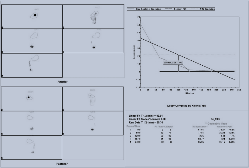
CT angiogram findings indicated severe MALS affecting the right kidney and coeliac artery, with compression, stenosis, and arterial collaterals. Upper GI endoscopy showed grade B distal esophagitis/antral gastritis. 99m Tc Sulphur Colloid scintigraphy revealed rapid gastric emptying, with a notably faster half-emptying time compared to the normal range.
DISCUSSION: Published literature and discussion surrounding MALS revolves around diagnostic challenges and potential etiologies, with theories suggesting either nerve dysfunction or mesenteric ischemia. Advanced imaging techniques like CT angiography play a crucial role in visualizing characteristic artery compression. The observation of rapid gastric emptying suggests potential involvement of reflux or gastritis. Moreover, the interplay between MALS and Gilbert syndrome, alongside the possibility of nerve plexus compression leading to delayed gastric emptying, underscores the complexity of this condition.
This case emphasizes the need for comprehensive investigation and management strategies tailored to individual patients with MALS. Treatment options may include surgical interventions such as laparoscopic or robotic median arcuate ligament release, aimed at alleviating symptoms and improving quality of life.

Fig A: shows rapid gastric emptying on 99m Tc sulphur colloid imaging after administration of 1mCi of radionuclide mixed with idli and delayed static imaging at 1 hourly intervals till 4hrs; image also reveals a raw data T1/2 of 35 minutes and more than 70% emptying in 1hr
References
- Lu, L. Y., Eastment, J. G., & Sivakumaran, Y. (2024). Median Arcuate Ligament Syndrome (MALS) in Hepato-Pancreato-Biliary Surgery: A Narrative Review and Proposed Management Algorithm. Journal of Clinical Medicine, 13(9), 2598.
- Balaban, D. H., Chen, J., Lin, Z., Tribble, C. G., & McCallum, R. W. (1997). Median arcuate ligament syndrome: a possible cause of idiopathic gastroparesis. American Journal of Gastroenterology, 92(3), 519-523.
- Reuter, S. R., & Bernstein, E. F. (1973). The anatomic basis for respiratory variation in median arcuate ligament compression of the celiac artery. Surgery, 73(3), 381-385.
- Jimenez, J. C., Harlander-Locke, M., & Dutson, E. P. (2012). Open and laparoscopic treatment of median arcuate ligament syndrome. Journal of vascular surgery, 56(3), 869-873.
- Tembey, R. A., Bajaj, A. S., k Wagle, P., & Ansari, A. S. (2015). Real-time ultrasound: key factor in identifying celiac artery compression syndrome. Indian Journal of Radiology and Imaging, 25(02), 202-205.
- Gruber, H., Loizides, A., Peer, S., & Gruber, I. (2012). Ultrasound of the median arcuate ligament syndrome: a new approach to diagnosis. Medical ultrasonography, 14(1), 5-9.
- Huynh, D. T., Shamash, K., Burch, M., Phillips, E., Cunneen, S., Van Allan, R. J., & Shouhed, D. (2019). Median arcuate ligament syndrome and its associated conditions. The American Surgeon, 85(10), 1162-1165.


