
Research Article | DOI: https://doi.org/10.58489/2833-0943/014
1 College of Veterinary Medicine, Department of Veterinary Public Health, Haramaya University, Eastern Hararghe, Ethiopia.
2 Department of Veterinary Medicine, Haramaya Woreda Veterinary Clinic, Eastern Hararghe, Ethiopia
3 Department of Veterinary Medicine, Gurawa Woreda Dogu Veterinary Clinic, Eastern Hararghe, Ethiopia
4 Department of Veterinary Medicine, Bedeno Woreda Furda Veterinary Clinic, Eastern Hararghe, Ethiopia
*Corresponding Author: Mohammed Kemal Mustefa Ame*
Citation: Mohammed Kemal Mustefa Ame,(2023)Assessment of Camel Mange Mite Prevalence in Kumbi Woreda, Eastern Harergae, Ethiopia. Journal of Pesticide Science and Pest Control 2(1). DOI: 10.58489/2833-0943/014
Copyright: © 2023 Mohammed Kemal Mustefa Ame, this is an open access article distributed under the Creative
Commons Attribution License, which permits unrestricted use, distribution, and reproduction in any medium,
provided the original work is properly cited.
Received: 01 April 2023 | Accepted: 12 April 2023 | Published: 14 April 2023
Keywords: Camel, Kumbi, Mange mite, Sarcoptes, Skin scraping
Camel mange, an exceedingly infectious ectoparasitism brought on by the parasite mite Sarcoptes scabiei and spread through direct or indirect contact, is one of the most significant parasitic disorders afflicting camels in Ethiopia. Economically significant infectious camel illness called camel mange affects the health and production of these animals. In order to ascertain the prevalence of camel mange mites and related risk factors in Kumbi woreda in Eastern Hararghe, Ethiopia, a cross-sectional study was carried out between May and November 2019. A total of 384 animals were randomly chosen for this investigation, and 96 camels tested positive for mange mite infestation. Skin scrapings were used to recover mange from suspicious lesions, which were then inspected under a microscope. As a result, 25% of camels were discovered to have mange mites. In every skin scraping sample taken from the suspected mange mite lesions, only Sarcoptes scabiei var. cameli mite species was found. PA, sex, age, physical condition status, and herd size were thought to be risk factors. Only the herd size and body condition shows a statistically significant difference in the prevalence of mange mites (P 0.05), whereas the PA, sex, and age did not exhibit any significant differences in mange infestation. According to this study, mange mites are present in camel papulations in the Kumbi woreda, which may negatively impact the health and productivity of the camels. To lessen the impact of mange mite infestation on camel husbandry, more emphasis should be placed on improving the management system, along with additional research and control measures.
Most of Africa's resource-poor farmers live in arid and semi-arid regions, where camels play a significant socioeconomic role (Wilson, 1984; Guliye et al., 2007; Mehari et al., 2007). There are an estimated 25.89 million camels in the world, spread over 47 nations. Eastern and northern Africa is home to the majority of the world's camels, with the remaining 15% living in nations in the Indian subcontinent and the Middle East (FAO, 2008). There has been a steady increase in the number of camels while the ongoing drought in the Horn of Africa is inflicting enormous losses in other livestock species, due to the camel's well-known capacity to produce in areas of drought when other animals rarely survive (Dioli and Stimmelmayr, 1992).
One of the nations with the highest camel densities in the world is Ethiopia. It comes in third place in Africa behind Sudan and Somalia. Its spread is greatly influenced by how well it can tolerate intense heat and desiccation. The subtropical arid parts of Asia and Africa make up the camel's natural range (Wilson et al., 1990). The main regions where camel herding is extensively practiced are the eastern and southern regions of the nation, including Afar, Somali, and Borena. Dromedaries undoubtedly sustain the livelihood of pastoral populations in this region (Tefera, 2009).
Camels are a valuable source of meat, milk, and dung, which is used in fires. In addition to being utilized for transportation, camels are mostly sold to Egypt and Sudan and are also killed for use in ceremonial meat eating (Dirie and Abdurahman, 2003). Even though camels have many socioeconomic benefits and are the ideal domestic animal species in the constantly changing environment, researchers and development planners have so far ignored this fact (Bekele, 2010). It has been determined that a number of endo and ectoparasites are the main issues influencing the health, productivity, and performance of camels (Anwar and Khan, 1998; Parsani et al., 2008; Bekele, 2010).
Although camels were long believed to be immune to many disease-causing factors, it has since been established that camels are just as susceptible as other livestock, if not more so, to the common infections that cause disease in other animal species (Abbas and Omar, 2005). Despite the importance of dromedaries in the difficult and hostile semi-arid and desert environments, their full utilization has been hindered by pathogenic infections, inadequate diet, outdated management methods, and a lack of veterinary services (Bekele, 2002).
Camel mange, an exceedingly infectious ectoparasitism brought on by the parasite mite Sarcoptes scabiei and spread through direct or indirect contact, is one of the most significant parasitic disorders afflicting camels in Ethiopia (Kumar et al., 1992). The productivity and growth of camel herds are severely hampered by the slow reproductive cycle, high calf mortality, and health issues. Productivity and performance are significantly hampered by camel ectoparasites (mites, ticks, and insects), which are also capable of spreading illness (Dioli and Stimmelmayr, 1992).
The economic worth of a mange-infested animal stems from the animal's lower body weight, the cost of treatment, the animal's skin deteriorating owing to skin perforation and acute itching, and occasionally dying in untreated or young animals (Wilson, 2008).
Moreover, the mange mite has significant zoonotic and public health implications (Singh, 2005; Wilson, 2008). For any attempt at prevention and control of the illness in question, knowledge about the prevalence of the diseases along with related risk factors as part of the epidemiology of the disease is essential. Despite several studies being conducted on other animal species, there hasn't been any research on the frequency of and risk factors for camel mange in Kumbi woreda. Therefore, the objectives of this study were:
The study was conducted in three purposefully selected peasant associations in Hararghe district of Kumbi woreda, Eastern Ethiopia between the period of May and November 2019. Study area was located 814 kilometers east of Addis Ababa. It is located 308 km away from the city of Harar. It is bordered by Gola Oda and Mayu Muluke woreda to the North, Burqua Dhintu to the West, Somali regional state to the East and South direction. The annual maximum and minimum temperatures are 28 and 16°C, respectively. The mean annual rainfall is 1300 mm and agro-climatic condition of the area is semi-arid and arid.There are two rainy seasons in the area, meher (June-September), used for crop production, pasture and water harvest and the short belg rains (February-May), mainly used for land preparation, planting of long cycle crops collected after the meher rains, small scale production, and improving water and pastures. Total livestock population of this district are 696,440; of which 206678 Cattle, 191444 goats, 140238 sheep, 19152 donkeys, 43 mules, 131857camels and 4488 poultry and 2540 Bee hives (KWVS, 2018).
The study animals were indigenous breeds of one humped camel (camelus dromedaries) reared under pastoral management system in free grazing, and usually mixed with livestock from other districts, the animals move from feed shortage area to feed abundant areas especially during drought season. Camels of all age categories and both sexes were included in the study.
A cross sectional study was used to estimate the prevalence and associated risk factors for the occurrence of camel mange in the study area.
The Peasant associations located in Kumbi woreda were selected purposively, based on the accessibility study population, willingness of the camel holder, considering their settlements, road accessibility and transport. The study animals were sampled by simple random sampling method.
Sample size determination was based on 50% expected prevalence, confidence interval 95% and desired level of precision 5%.The total number of samples required for this study were calculated based on the following formula (Thrusfield, 1995).
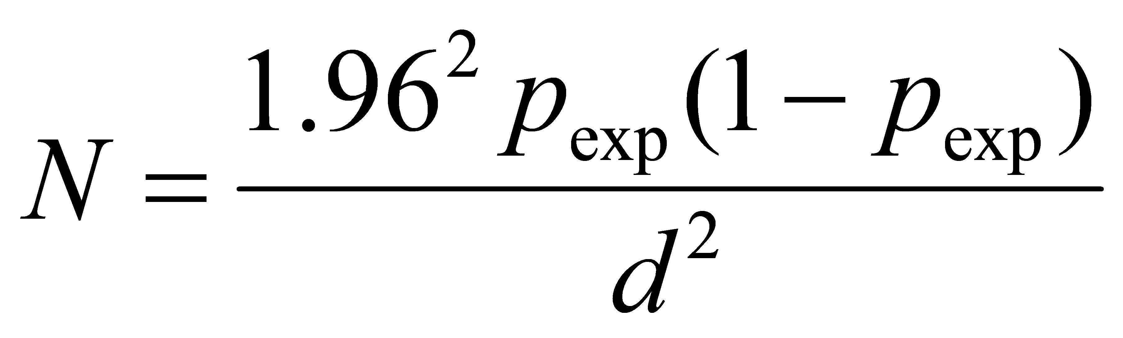
When: N= required sample size; P=expected prevalence; d=desired level of precision. There was no previously documented camel mange mite in the study area. Accordingly, the minimum samples sizes required for this study were 384.
Skin scrapings of 384 camels with considering of peasant association, age, sex, body condition, and herd size, was taken from different camel populations in Kumbi woreda. Age of studied camels was categorized into <3>4 years as an adult camel. The age of the sampled animals was determined by dental eruption according to (FAO, 1994). The body condition score (BCS) of sampled camels was evaluated by looking the back and flank then categorized as good, medium and poor according to Faye et al., (2001). Herd sizes as small (less than twenty), as medium (between twenty and forty) and large (greater than forty) were determined according to classification of (Bekele et al., 2012). After selection of animals, each camel was restrained properly and the hairs were shaved using scalpel blade from the edges of the lesions till blood oozes out of the capillary (Soulsby, 1982).
Skin scrapings from suspected cases of mange were collected in labelled Petri-dishes and preserved in 10% formalin and taken to laboratory and 10% potassium hydroxide (KOH) was added to digest or clean the scraped material of skin, hair, and other debris so that mites released from scabs and crusts before examination following procedures indicated by (Soulsby, 1982). All scraped tissues were carefully placed on microscopic slide for microscopic examination (10 x or 40 x magnifications) and identification of the mange mite species based on the morphological characteristics described by (Urquhart et al., 1996).
Microsoft excel spread sheet program was used to store all the data and Statistical Package for Social Sciences (SPSS) version 22.00 software was used to analyze the data. Prevalence of mange mites was computed as the number of each sample items positive for mange divided by total number of the samples examined. Chi-square (χ2) was used to test the presence of association between variables. When P value was less than 0.05, the presence of significance difference was considered.
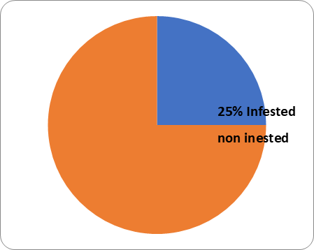
The overall prevalence of camel mange mites in this study was found to be 25% (96/384). In this study, only sarcoptes scabiei var. cameli was identified as the only mite species in all skin scraping samples collected from the suspected lesions.
In the study area, Urgo and Ija-Godda were found with slightly higher prevalence followed by Kara-Balchi having 28.1%, 24.6% and 22.1% respectively. There is no statistically significant difference in the occurrence of camel mange among peasant associations (p > 0.05) (Table 1).
Table 1: Prevalence of camel mange among peasant associations
| PA | Sample Examined | Positive | Prevalence (%) | χ2 | P-value | ||
| Urgo | 128 | 36 | 28.1% | 1.212 | 0.545 | ||
| Kara-Balchi | 122 | 27 | 22.1% | ||||
| Ija-Godda | 134 | 33 | 24.6% | ||||
| Total | 384 | 96 | 25% | ||||
The sex wise prevalence of camel mange mite in the study area was revealed 26.4%and22.8% in female and male respectively. There was no statistically significance difference between the two sex groups(P>0.05)(Table 2).
Table 2 .Prevalence of camel mange between sexes
| Sex | Sample Examined | Positive | Prevalence (%) | χ2 | P-value |
| Male | 149 | 34 | 22.8% | 0.618 | 0.432 |
| Female | 235 | 62 | 26.4% | ||
| Total | 384 | 96 | 25% |

The current study showed a higher prevalence of mange mites infestation in young age group of camels
(31.8%), than adult age group of camels (23.1%), and the difference was not statistically significant(p>0.05)(Table3).
Table 3. Prevalence of camel mange between ages
| Age | Sample Examined | Positive | Prevalence (%) | χ2 | P-value |
| Young | 85 | 27 | 31.8% | 2.664 | 0.103 |
| Adult | 299 | 69 | 23.1% | ||
| Total | 384 | 96 | 25% |
In this study, the prevalence of camel mange was higher in camels with poor body condition (33.1%), followed by medium (22.9%) and good body conditioned camels (11.3%) and the variation was statistically significant (P<0>
Table 4 .Prevalence of camel mange among body condition scores
| BCS | Sample Examined | Positive | Prevalence (%) | χ2 | P-value |
| Poor | 160 | 53 | 33.1% | 13.142 | 0.001 |
| Medium | 153 | 35 | 22.9% | ||
| Good | 71 | 8 | 11.3% | ||
| Total | 384 | 96 | 25% |
The analysis result of this study revealed that small (<20>40) herd sizes had the prevalence of 17.5%, 27.1% and 34.3% respectively and the variation was statistically significant in their prevalence (P<0>
Table 5.Prevalence of camel mange among herd sizes
| Herd Size | Sample Examined | Positive | Prevalence (%) | χ2 | P-value |
| Small | 137 | 24 | 17.5% | 7.733 | 0.021 |
| Medium | 177 | 48 | 27.1% | ||
| Large | 70 | 24 | 34.3% | ||
| Total | 384 | 96 | 25% |
Current study showed an overall prevalence of 25% mange mite infestation among camelherds. This result was in line with the various works done by Bekele et al. (2012) in Borana, Southern Ethiopia, Abebe (2001) and Teka et al. (2017) in Eastern Ethiopia who reported a prevalence of 25.9%, 27.8% and 32.4% respectively. However, this finding was higher than the reports of Awol et al. (2014) in Azebu district, northern Ethiopia, Zahid et al. (2015) in Punjab, Pakistan, Dinka et al. (2010) eastern Ethiopia, Lawal et al. (2007) Sokoto, Nigeria and Chaudhry et al. (2014) Cholistan, Pakistan whose results were 16.7%,11.28%, 10.7%, 3.5% and 3.14% respectively. These discrepancies in the prevalence of camel mange mite among different studies could be due to variations in environment, study seasons, level of awareness of the community with regard to methods of transmission and control and animal husbandry and managements.
Sarcoptes scabiei var. cameli was identified as the only mite species in all scrapings collected from suspected skin lesions. The same findings have been encountered by numerous authors like (Bekele et al., 2012), (Awol et al., 2014), (Zahid et al., 2015) and (Teka et al., 2017).Even though both sarcoptic and chorioptic mange mites have been reported, Sarcoptic mange caused by Sarcoptes scabiei var. cameli is by far the most common, contagious and serious condition in camels (Pegram and Heggins, 1992; Parsani, 2008).
There was no significant variation (P>0.05) in the prevalence of camel mange mite infestation between the peasant associations, sexes, and age. This finding was in general agreement with reports of (Teka et al., 2015) from eastern Ethiopia and Bekele et al. (2012) in Borana, southern Ethiopia. This relation might be due to similarities in management and the availability of same veterinary services as well as micro climatic condition of the study area. But there was significant difference (P<0>et al. (2012) in Borana, Southern Ethiopia and Awol et al. (2014) in Azebu district, Northern Ethiopia; it could be due to a variation in environment, study seasons and management practices.
On the other hand, the increment of prevalence of female animals than that of male animals in the study might be due to hormonal influences i.e. the higher level of prolactin and progesterone hormones could make the females more susceptible to any infection. Additionally, pregnancy and lactation stress could also aggravate the susceptibility of the female camels to infections. Furthermore, the breeding behaviour of mange infected males could also be attributed to the transfer of the disease to a number of females (Lioyd, 1983).
Higher prevalence of mange mite was recorded in the young animals than the old one. This finding is in agreement with the others work (Dinka et al., 2010; Ashraf et al., 2014; Awol et al.,2014). The age of camels might be important factors in mange infestation, in which both very young and very old camels are particularly susceptible. The increased prevalence of mite infestation in young camels with 4 years of age than the rest age groups could be probably reflecting lowered body's defences of young animals. Furthermore, close interaction of the sucklers with infested lactating females could also be another factor which makes them more liable to the disease, leading to a higher prevalence in this age group.
With regards to herd size, the present study shown an escalation in the prevalence of Sarcoptes scabiei in herds with larger size which is most probably to the fact that camels from large herd sizes are more prone to be exposed to diseased animals supporting the contagious nature of mite infestation and contacts during herding, housing and interactions at watering points and auction marts favours the establishment and spread of mite infestation.
This study was done to find out how common camel mange mites are and what risk factors they have in Kumbi woreda. As a result, 96 (25%) of the 384 camels that were screened for the presence of the parasite were confirmed to be positive. The predominant cause of mange in camels in the research region is Sarcoptes scabiei var. cameli. According to the present findings, among the risk factors considered throughout the study, camel mange infestation was more prevalent in animals with poor physical condition and big herd sizes.
Due to a lack of food, the camel population in the area is more likely to come into close contact with other animals at available communal watering holes, which hastens the development and spread of mite infection. This study demonstrated that camels have large mange mite populations, which may have a negative impact on the health and productivity of these animals. These findings' conclusions led to the following recommendations being made: