
Research Article | DOI: https://doi.org/10.58489/2836-5127/003
Advisor doctor and expert trainer, Baghdad Medical City and the National Training and Development Center, Iraqi Ministry of Health, Baghdad, Iraq
*Corresponding Author: Aamir Jalal Al-Mosawi
Citation: Aamir Jalal Al-Mosawi, (2023). Bilateral ectopic pelvic kidneys: The novel association with liver hemangioma. Journal of Radiology Research and Diagnostic Imaging. 2(1). DOI: 10.58489/2836-5127/003
Copyright: © 2023, Aamir Jalal Al-Mosawi, this is an open access article distributed under the Creative Commons Attribution License, which permits unrestricted use, distribution, and reproduction in any medium, provided the original work is properly cited.
Received: 10 December 2022 | Accepted: 23 December 2022 | Published: 09 January 2023
Keywords: Bilateral ectopic pelvic kidneys, hepatic hemangioma.
Background: Renal ectopia is a congenital abnormality in which one or both kidneys are located in an unusual position because of failure of normal ascend from its origin in the true pelvis. Several types of renal ectopia with or without fusion or other associated renal and abdominal visceral abnormalities have been reported. We have previously reported the case forty-one of crossed unfused renal ectopia, and the aim of this paper is to report the association of bilateral ectopic pelvic kidneys with liver hemangioma.
Patients and methods: The case of a woman in her mid-forties with renal and hepatic abnormalities on pelvic and abdominal ultrasound was studied.
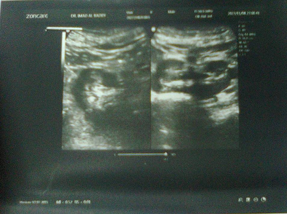
Results: A woman in her mid-forties was having recurrent lower abdominal pain or discomfort and recurrent urinary tract infection. Ultrasound showed that both kidneys were ectopic and were located in the pelvic cavity. Both kidneys were normal in size, texture, parenchymal thickness, and had normal cortico-modularly differentiation. There was mild splitting of the pelvi-calyceal system suggesting infection. Both ureters were normal. The liver was normal in size with homogenous texture, but there was a right lobe well-defined echogenic soft tissue mass, hemangioma. Renal function tests showed normal findings.
Conclusion: Bilateral ectopic pelvic kidneys is a rare congenital condition that has not been reported to occur in association with hepatic hemangioma. This paper reports the novel association of bilateral ectopic pelvic kidneys with hepatic hemangioma.
Renal ectopia is congenital abnormality in which one or both kidneys are located in an unusual position because of failure of normal ascend from its origin in the true pelvis. Several types of renal ectopia with or without with or without fusion or other associated renal and abdominal visceral abnormalities have been reported since the early report of Polk in I882 [1, 2, 3]. We have previously reported the case forty-one of crossed unfused renal ectopia [1], and the aim of this paper is to report the association of bilateral ectopic pelvic kidneys with liver hemangioma.
The case of a woman in her mid-forties with renal and hepatic abnormalities on pelvic and abdominal ultrasound was studied.
A woman in her mid-forties was found to have renal and hepatic abnormalities on pelvic and abdominal ultrasound during the evaluation after treatment for urinary tract infection. She was having recurrent lower abdominal pain or discomfort and recurrent urinary tract infection. Ultrasound (Figure-1) showed that both kidneys were ectopic, and were located in the pelvic cavity. Both kidneys were normal in size, texture, parenchymal thickness, and had normal cortico-modularly differentiation. There was mild splitting of the pelvi-calyceal system suggesting infection. Both ureters were normal. The liver was normal in size with homogenous texture with a right lobe well-defined echogenic soft tissue mass, hemangioma. She had normal blood pressure and renal function tests showed normal findings.
As early as 1931 Howard L Tolson emphasized that ectopic kidneys can be structurally and functionally normal and remain asymptomatic or associated with mild dull pain and urinary tract infections [4].
Boujnah et al (1989) from Tunisia emphasized that bilateral pelvic ectopic kidneys is a rare congenital condition, and they reported 50 cases of pelvic ectopic kidneys observed during 12 years. 47 patients had unilateral pelvic ectopic kidney, two patients had bilateral pelvic ectopic kidneys, and one patient had pelvic ectopic kidney solitary pelvic ectopic kidney. Eighteen patients had healthy ectopic kidneys. Thirty-two patients had diseased ectopic kidney including twenty-one patients with renal stones and seven patients with uretero-pelvic junction disease.
Boujnah et al from Tunisia emphasized the diagnostic value of renal ultrasound which can help in avoiding other useless and expensive investigations [5].
Hirano and colleagues (1992) reported the incidental detection of bilateral ectopic pelvic kidneys by radioisotope angiography [6].
Alonso Domínguez (1996) also emphasized the rarity of bilateral pelvic ectopic kidneys and reported a case with an unusual presentation [7].
Gokalp and colleagues (2010) reported a case with bilateral ectopic kidneys associated with vascular anomaly and hypertension and renal dysfunction [8].
Hemangiomas including liver hemangiomas are benign vascular tumors that are generally observed during infancy. Liver hemangioma is the most common benign tumor of the liver [8, 9]. Brodsky et al (1987) reported that during the performance of high-resolution real-time abdominal sonography, hemangioma small echogenic hepatic masses are frequently discovered [10].
Lipman and Tumeh (1990) from Boston, Massachusetts emphasized that the diagnosis of small liver hemangiomas, less than 3 cm can be made with abdominal ultrasound study [11].
Al-Durazi et al (2003) from Bahrain reported the incidental finding of a case of hepatic hemangioma while performing a routine renal and pelvic ultrasound in patients with urinary retention associated with benign prostatic hyperplasia [12].
Mungovan and colleagues (1994) emphasized that the size of the majority of hepatic hemangiomas don’t increase in size for months and years, and an increase in size demands further evaluation [13].
Bilateral ectopic pelvic kidneys are a rare congenital condition that has not been reported to occur in association with hepatic hemangioma. This paper reports the novel association of bilateral ectopic pelvic kidneys with hepatic hemangioma.