
Research Article | DOI: https://doi.org/10.58489/2836-5038/002
Grodno State Medical University, Grodno, Republic of Belarus.
*Corresponding Author: E.I. Bon
Citation: E.I. Bon, N. E. Maksimovich, S.M. Zimatkin, O.B. Ostrovskaya, V.Yu Smirnov, M.A. Nosovich, K.A. Khrapovitskaya, Portonenko A.M., (2022). Changes in the Organelles of Neurons in The Parietal Cortex and Hippocampus in Incomplete Cerebral Ischemia. International Journal of Stem cells and Medicine. 1(2). DOI: 10.58489/2836-5038/002.
Copyright: © 2022 E.I. Bon, this is an open access article distributed under the Creative Commons Attribution License, which permits unrestricted use, distribution, and reproduction in any medium, provided the original work is properly cited.
Received: 02 November 2022 | Accepted: 29 November 2022 | Published: 23 December 2022
Keywords: neurons, parietal cortex, hippocampus, ischemia
The ultrastructural characteristics of neuronal organelles are important indicators of the degree of brain damage during ischemic exposure, which necessitates studying changes in the ultrastructure of brain neurons.
Aim: To study the nature of disorders of brain neurons in case of its partial ischemia at the ultrastructural level.
Materials and Methods: Experiments were performed on 12 males weighing 260±20 g. Partial cerebral ischemia was modeled by ligation of one common carotid artery on the right. The material was taken 1 hour after the operation. The study group included 8 males, the control group consisted of 4 falsely operated rats of the same sex and weight
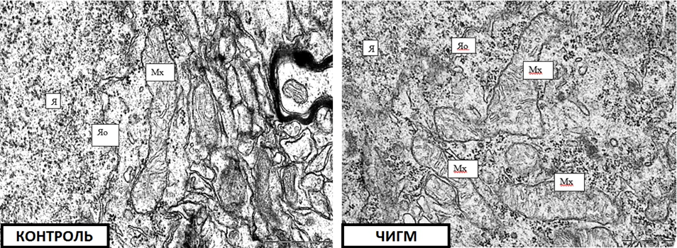
Results: An electron microscopic study showed that the size and shape of mitochondria of neurons in the parietal and hippocampus of rats with partial cerebral ischemia do not differ from the size and shapes of mitochondria of neurons in the parietal cortex and hippocampus of rats in the control group (p>0.05), except for a lower density of crista. in mitochondria of neurons of the parietal cortex by 18%, p<0.05.
Conclusions Thus, the ultrastructure of neurons in partial cerebral ischemia is generally similar to that in the control group, which may be due to compensation of blood flow in the circle of Willis.
With cerebral ischemia (BCI), a chain of pathogenetic disorders develops in its structures, among which one of the leading ones is energy deficiency, which leads to the development of cellular pathology due to disturbances in homeostasis, enzyme activity, membrane integrity and energy pumps. Under conditions of cerebral ischemia, the mechanisms of synaptic transmission are selectively disrupted, which contributes to impaired local blood flow autoregulation, the development of vasospasm, increased platelet aggregation, and the development of intravascular stasis, aggravating hypoxia and increasing energy deficiency. The functioning of enzymes, including sodium-potassium ATPase, is disrupted, leading to an imbalance of ions and cerebral edema [1,2,3,4].
The ultrastructural characteristics of neuronal organelles are important indicators of the degree of brain damage during ischemic exposure, reflecting the severity of compensation, which necessitates the study of changes in the ultrastructure of brain neurons.
According to the literature, during BCI in the cytoplasm of neurons, swelling of mitochondria and destruction of their cristae, expansion of cisterns of the endoplasmic reticulum and the Golgi complex, and an increase in the number of free ribosomes, which form extensive clusters in the cytoplasm, are observed. The total number of lysosomes increases, their sizes increase. However, there are no data on the severity of these disorders depending on the type of ischemic injury and its severity [5-12].
To study the nature of disorders of brain neurons in case of its partial ischemia at the ultrastructural level.
The experiments were carried out on 12 male outbred white rats weighing 260±20 g in compliance with the requirements of the Directive of the European Parliament and of the Council No. 2010/63/EU of September 22, 2010 on the protection of animals used for scientific purposes.
Modeling of BCI was carried out under intravenous thiopental anesthesia (40-50 mg/kg).
The study used a model of partial (PCI) cerebral ischemia. Partial cerebral ischemia or PCI was modeled by ligation of one CCA on the right. The material was taken 1 hour after the operation.
The study group included 8 males, the control group consisted of 4 falsely operated rats of the same sex and weight.
Electron microscopic studies were performed in the parietal cortex and hippocampus of the brains of rats.
Immediately after decapitation and quick extraction of the brain, parts of the parietal cortex and hippocampus were cut with a blade and placed in 1% osmium fixative in Millonig's buffer (pH=7.4) for 2 hours at 4°C.
Then the sections were washed in a mixture of Millonig's buffer (20 ml) and sucrose (900 mg), dehydrated in alcohols of increasing concentration, a mixture of alcohol and acetone, and pure acetone; passed through a mixture of resins (araldite M + araldite H + dibutyl phthalate + DMR-30) and acetone and placed in a mixture of resins.
Semithin sections (about 350 nm thick) were made on an MT-7000 ultramicrotome (RMC, USA) and stained with methylene blue. Next, sections of the inner pyramidal layer of the parietal cortex and the pyramidal layer of the field CA1 of the hippocampus, necessary for study, were cut out with a blade.
Ultrathin sections (thickness about 35 nm) were made on the same ultramicrotome, collected on supporting grids, contrasted with uranium acetate and lead citrate. To do this, the meshes with sections were dipped into a drop of uranyl acetate and kept for 20 minutes in the dark at room temperature, then washed in 3 portions of bidistilled water for 5 seconds and contrasted with lead citrate for 8 minutes, washed in 3 portions of bidistilled water for 5 seconds.
The resulting preparations were studied under a JEM-1011 electron microscope (JEOL, Japan) and photographed with an Olympus MegaView III digital camera (Olympus Soft Imaging Solutions, Germany).
Morphometry of ultrastructures was performed using the Image Warp image processing program (Bit Flow, USA), for which mitochondria, the Golgi complex, the granular endoplasmic reticulum, ribosomes, and lysosomes were circled on a computer monitor. Density, size, shape of organelles, density and length of mitochondrial cristae, sizes of lysosomes and their density, the number of endoplasmic reticulum bound and free ribosomes were measured, and their ratio coefficient was calculated.
To prevent a systematic measurement error, brain samples from the compared control and experimental groups of animals were studied under the same conditions.
As a result of the research, quantitative continuous data were obtained. Since the experiment used small samples that had a non-normal distribution, the analysis was performed by nonparametric statistics using the licensed computer program Statistica 10.0 for Windows (StatSoft, Inc., USA). Data are presented as Me (LQ; UQ), where Me is the median, LQ is the lower quartile value; UQ is the value of the upper quartile. Differences between groups were considered significant at p<0>
An electron microscopic study showed that the size and shape of the mitochondria of the parietal cortex and hippocampus neurons of rats with CCI did not differ from the size and shape of the mitochondria of the parietal cortex and hippocampus neurons of control rats (p>0.05), with the exception of a lower density of cristae in the mitochondria of the parietal neurons. bark by 18%, (p<0>
Table 1: Parameters of ultramicroscopic morphometry of neuron organelles in the parietal cortex and hippocampus of rats with partial cerebral ischemia, Me (LQ; UQ).
Index | Parietal cortex | Hippocampus | |||
Control | PCI 1 hour | Control | PCI 1 hour | ||
Mitochondria | density | 1,8(1,7;2,2) | 1,9(1,6;2,3) | 2,1(1,7;2,2) | 2,2(1,6;2,4) |
area, µm2 | 0,26 (0,17;0,37) | 0,27 (0,18;0,33) | 0,21 (0,17;0,26) | 0,22 (0,18;0,25) | |
Form factor, unit | 0,63(0,61;0,72) | 0,69(0,60;0,75) | 0,71(0,60;0,75)* | 0,76(0,66;0,78) | |
Elongation factor, units | 3,8(3,5;4,1) | 3,7(3,5;4,1) | 2,1(1,9;2,5) | 2,0(1,9;2,2) | |
mitochondrial crist density | 76(71;82) | 62(58;67)* | 62(59;72) | 68(60;78) | |
length of mitochondrial cristae / µm2 | 12(10;15) | 10(9;12) | 13(12;18) | 13(10;15) | |
Ribosomes | amount /µm2 | 20,9(19,3;22,7) | 22,3(19,1;24,2) | 20,0(18,1;22,8) | 21,6(18,8;23,9) |
free / µm2 | 4,7(4,1;5,8) | 12,5(11,2;13,0) | 6,0(4,8;7,3) | 12,8(11,4;13,4) | |
bound /µm2 | 16,2(15,2;16,9) | 9,8(7,9;11,2) | 14,0(13,3;15,5) | 8,8(7,4;10,5) | |
ratio of bound and free ribosomes | 3,4 | 0,8* | 2,3 | 0,7* | |
Lysosomes
| density | 0,4(0,3;0,5) | 0,4(0,3;0,6) | 0,5(0,4;0,6) | 0,5(0,4;0,6) |
area, µm2 | 0,02(0,01;0,03) | 0,02(0,01;0,03) | 0,03(0,02;0,04) | 0,02(0,01;0,02) | |
Note: numerical values are presented as Me (LQ; UQ), * - p<0>
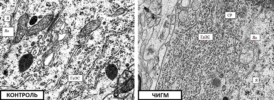
The size and shape of the Golgi complex and lysosomes did not differ from those in the control group either. However, in the cytoplasm of neurons of the parietal cortex and hippocampus of rats with PCI, an increase in the number of free ribosomes was noted - by 58% (p<0>
The coefficient of the ratio of bound and free ribosomes decreased from 3.4 in the control group to 0.8 in the parietal cortex (p<0>
According to the literature, during cerebral ischemia, a number of typical disorders of the ultrastructure of neurons occur, which are manifested by swelling of mitochondria and destruction of cisterns of the endoplasmic reticulum and the Golgi complex [1,2,7]. In PCI, such disturbances were insignificant and consisted only in the predominance of free ribosomes and the destruction of cristae in the parietal cortex, which is more sensitive to oxygen deficiency.
The mitochondria of rats with PCI did not differ in size and shape from the mitochondria of rats in the control group, with the exception of a lower density of mitochondrial cristae of parietal cortex neurons (Fig. 1).
However, in the cytoplasm of neurons in the parietal cortex and hippocampus of rats with PCI, an increase in the number of free ribosomes was noted (Fig. 2).
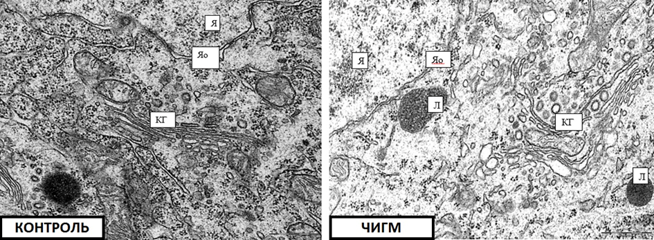
The average number and size of lysosomes, as well as CG (Fig. 3) did not differ from those in the control group.
Thus, the ultrastructure of neurons in partial cerebral ischemia is generally similar to that in the control group, which may be due to compensation of blood flow in the circle of Willis. However, the increase in the number of free ribosomes is a sign of a violation of protein biosynthesis in neurons in this model of CI, and a decrease in the density of neuron mitochondrial cristae in the parietal cortex indicates the occurrence of energy deficiency in this area of the brain, as it is more sensitive to oxygen deficiency.
The authors declare no conflict of interest.