
Research Article | DOI: https://doi.org/10.58489/2836-5003/003
Grodno State Medical University, Grodno, Republic of Belarus
*Corresponding Author: M.A. Feduto
Citation: M.A. Feduto, N.E. Maksimovich, S.M. Zimatkin, E.I. Bon, A.I. Grishchenko, U.A. Bakush, D.V. Gaiko, (2022). Comparative Characteristics of Morphological Changes in Neurons of The Parietal Cortex of Rats with Anoxia of Ischemic and Respiratory Genesis. Archives of Immunology Research and Therapy. 1(1). DOI: 10.58489/2836-5003/003.
Copyright: © 2022 M.A. Feduto, this is an open access article distributed under the Creative Commons Attribution License, which permits unrestricted use, distribution, and reproduction in any medium, provided the original work is properly cited.
Received: 09 December 2022 | Accepted: 16 December 2022 | Published: 23 December 2022
Keywords: anoxia, neurons, brain
Acute oxygen deficiency is the basis of a variety of pathological processes in many diseases and environmental factors. When studying the rat brain under conditions of its total ischemia and mechanical asphyxia, the presence of structural changes in both studied periods (after 30 and 60 minutes) was revealed: a decrease in the area and a change in shape (loss of sphericity and an increase in elongation) of cells, as well as a change in the degree of chromatophilia, which was manifested by a decrease in normochromic neurons with a simultaneous increase in hyperchromic shriveled neurons. At the same time, total cerebral ischemia led to more pronounced changes in the studied neurons of the parietal cortex, which was manifested by a more significant decrease in the area of neurons after 30 minutes of anoxia, while shape changes in the form of loss of sphericity and increased elongation were noted to a greater extent with mechanical asphyxia. These differences may be due to the preservation of cardiac activity during mechanical asphyxia for a short time.
Acute oxygen deficiency is the basis of various pathological processes in many diseases and environmental factors [2,3,5]. In particular, oxygen deficiency in the brain can occur most often as a result of hemodynamic disorders (prolonged spasm, thrombosis, embolism of the vessel) or the influence of an external mechanical factor (mechanical asphyxia) [7,10].
Anoxia as an extreme degree of oxygen starvation of brain cells, regardless of the causes, leads to its greatest damage. This is due to the complexity of the morphological structure and functions of the brain, as well as low tolerance to oxygen deficiency, which is determined by a high level of metabolism, lack of oxygen reserves and macroergic compounds [2,3,5].
The cerebral cortex deserves special attention, which is associated with the extreme importance in the vital activity of the body and the severity of the consequences that develop when it is damaged [2,5,6,7].
To date, the pathomorphological, pathophysiological and clinical aspects of global anoxia have been covered in sufficient detail in the literature [3,4,7]. The peculiarity of anoxia of respiratory genesis, in contrast to anoxia of ischemic genesis, is that the heartbeat persists for a certain time. However, there isno blood oxygenation. In this regard, it is advisable to carry out a comparative analysis of neuronal changes in the brain with its total ischemia and mechanical asphyxia.
The aim is to compare morphological changes in neurons of the parietal cortex of the rat brain during its anoxia caused by total ischemia and mechanical asphyxia.
The study was conducted on mongrel white rats (30 males, weight 240 ± 20 g), divided into 5 groups (n=6) in compliance with the requirements of Directive of the European Parliament and of the Council No. 2010/63/EU of 22.09.2010 on the protection of animals used for scientific purposes.
The use of rats as experimental animals is due to the similarity of angioarchitectonics and cytoarchitectonics of the brain in rats and humans.
The experiments were performed on 2 models of brain hypoxia: total ischemia and mechanical asphyxia. The control group consisted of falsely operated rats (group 1). Mechanical asphyxia was simulated by ligating the rat trachea for 30 minutes (group 2) and 60 minutes (group 3) [9]. Modeling of total cerebral ischemia was carried out by decapitation of rats with sampling 30 minutes (group 4) and 60 minutes (group 5) after decapitation [2].
The studies were carried out under intravenous anesthesia (sodium thiopental, 40 mg/kg). The brain was extracted and fixed in Carnoy's solution, after which frontal paraffin sections of the parietal cortex with a thickness of 7 microns were made and stained using the Nissl method [8]. The location of the parietal cortex was determined using a stereotactic atlas [12].
In each animal, 30 neurons of the fifth layer of the parietal cortex were studied to determine their size and shape [6,11]. Changes in the area and shape of neurons (form factor, elongation factor) were evaluated using the ImageWarp (Bitflow, USA). In histological preparations, various types of neurons were determined by the degree of staining of their cytoplasm (chromatophilia).
The obtained quantitative continuous data were processed using methods of nonparametric statistics, licensed computer program Statistica 10.0 for Windows (StatSoft, Inc., USA). The data is presented in the form Me (LQ; UQ), where Me is the median, LQ is the boundary of the lower quartile; UQ is the boundary of the upper quartile. The differences between the indicators of the control and experimental groups were considered significant at р<0>
In the control group, up to 95 percentage of the population of parietal cortex neurons were normochromic cells, and the remaining neurons were hypochromic (4 Percentage) and hyperchromic shriveled (1 percentage) cells.
In animals of the experimental groups, structural changes of neurons of the parietal cortex of the brain occurred, which manifested themselves in changes in the size, shape of neurons, and intensity of staining of their cytoplasm.
Hyperchromic shriveled neurons – elongated and polygonal neurons with intensely colored cytoplasm – prevailed in both studied time intervals in total ischemia and mechanical asphyxia, about 75percentage in the group of rats with 30–minute mechanical asphyxia (p<0 xss=removed> Thus, in total ischemia and mechanical asphyxia, unidirectional changes in chromatophilia were noted equally (p>0.05), which is manifested in a significant increase in hyperchromic shriveled neurons, which, as is known, are markers of acute oxygen deficiency of nervous tissue [2].
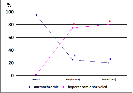
Dynamics of changes in the number of normochromic and hyperchromic shriveled neurons of the parietal cortex after 30 minutes and 60 minutes of mechanical asphyxia (MA) Note – * – the differences are reliable compared to the control (р<0>
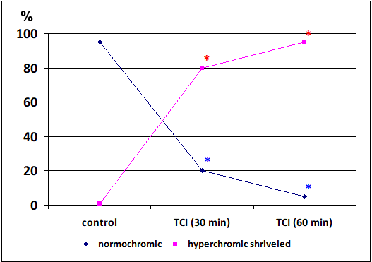
Dynamics of changes in the number of normochromic and hyperchromic shriveled neurons of the parietal cortex after 30 minutes and 60 minutes of total cerebral ischemia (TCI)
Note – * – the differences are reliable compared to the control (р<0>
There was no change in the area of neurons after 30 minutes of mechanical asphyxia (р>0,05). But at the same time, a change in the shape of neurons was noted: the form factor decreased by 23percentage (p<0 xss=removed>Fig. 3).
In rats with total cerebral ischemia, by 30 minutes of the ischemic period, the area of neurons decreased by 74percentage (p<0>percentage (p<0>percentage (p<0>
By 60 minutes of mechanical asphyxia, the area of neurons decreased by 35percentage (p<0>0.05).
By 60 minutes of total cerebral ischemia, there was no aggravation of changes in the size and shape of neurons (p>0.05).
A
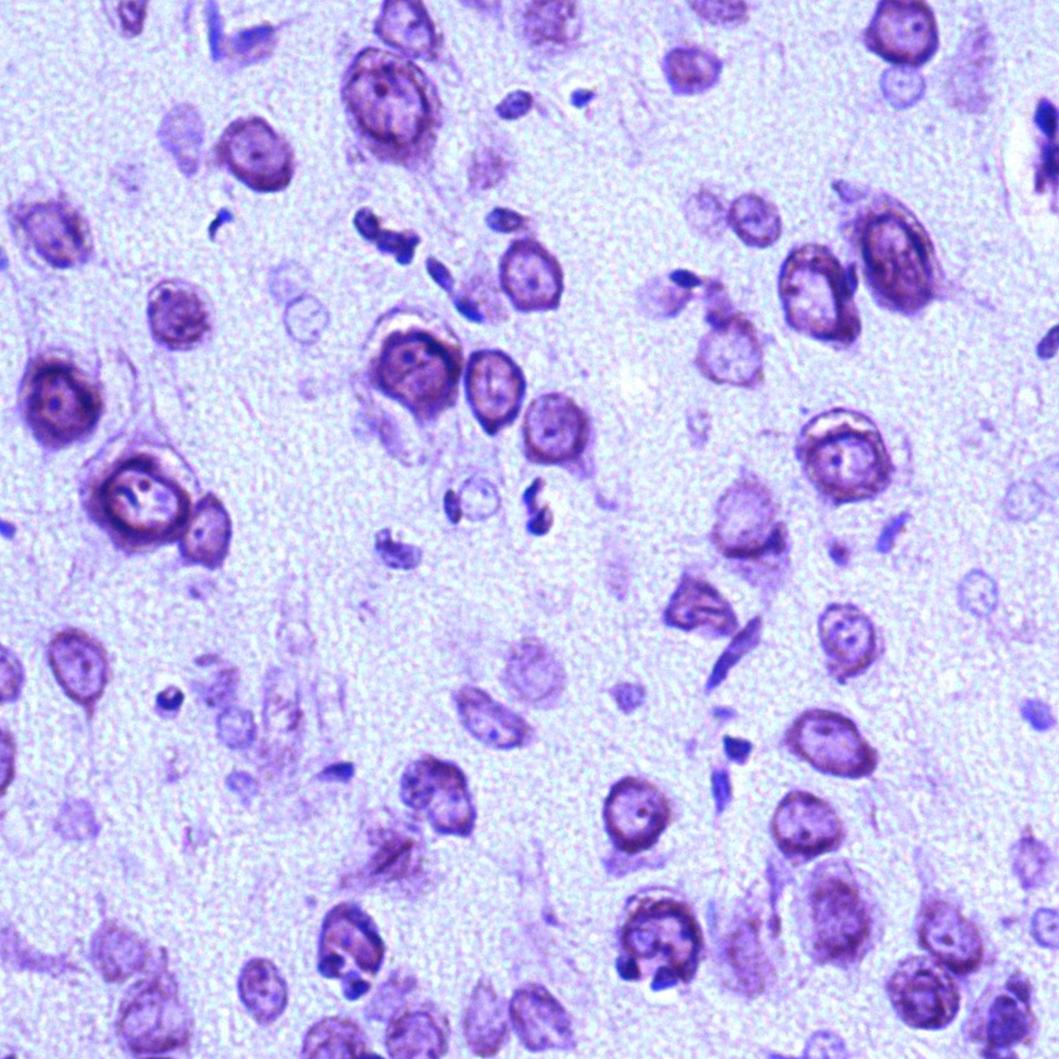 figure A
| B
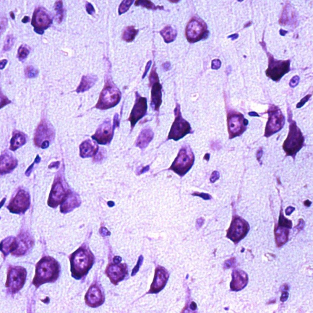 figure B
|
A | B |
C
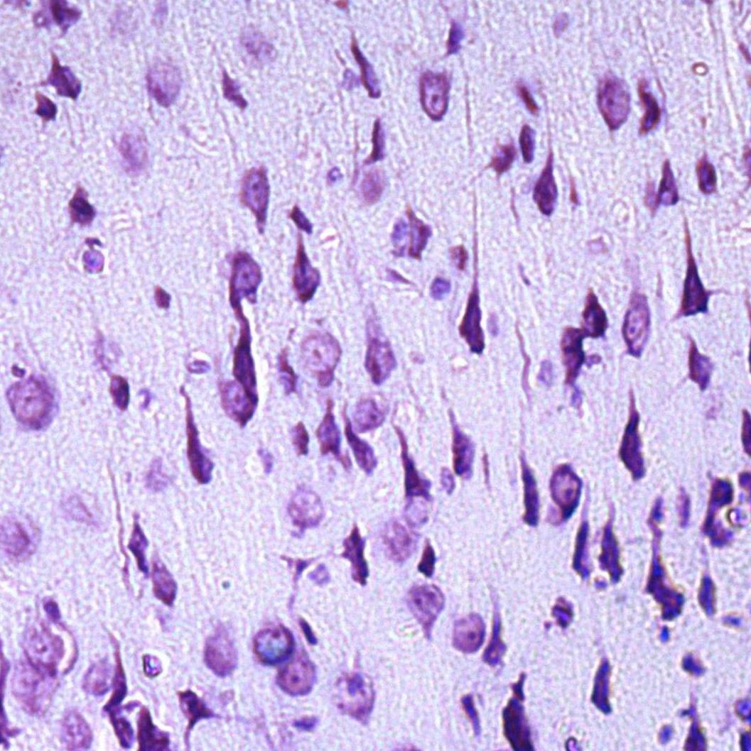 figure C
| D
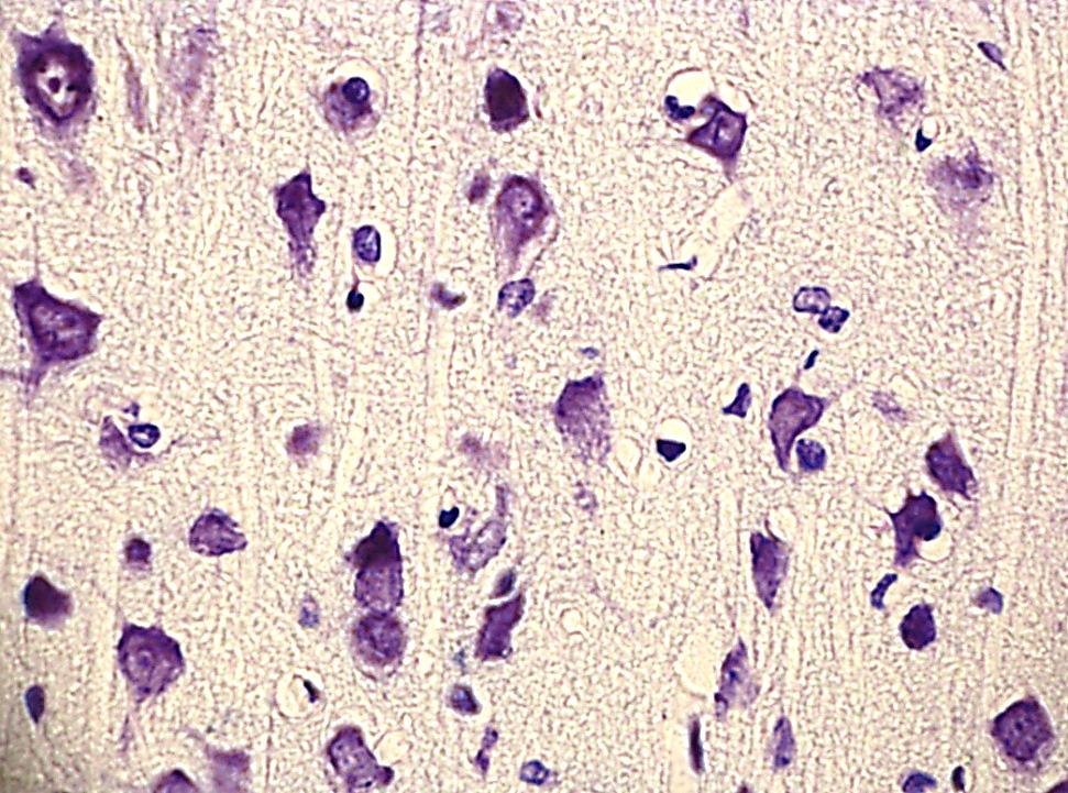 figure D
|
C | E |
E
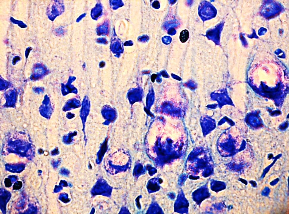 Figure 3. Neurons of the 5th layer of the parietal cortex of the rat brain. Digital microphotography. Stained by Nissl.
| |
A – the control group (predominance of normochromic neurons); B – after 30 minutes of mechanical asphyxia (predominance of hyperchromic shriveled neurons); C – after 60 minutes of mechanical asphyxia (predominance of hyperchromic shriveled neurons); Magnification x 20. D – after 30 minutes of total ischemia (predominance of hyperchromic shriveled neurons); E – after 60 minutes of total ischemia (predominance of hyperchromic shriveled neurons). Magnification x40.
So, the study of the effects of anoxia on neurons of the parietal cortex of the rat brain under conditions of its total ischemia and mechanical asphyxia revealed the presence of structural changes in both studied periods (after 30 and 60 minutes): reducing the area and changing the shape (loss of sphericity and increase in elongation) of cells, as well as a change in the degree of chromatophilia, which was manifested by a decrease in normochromic neurons with a simultaneous increase in hyperchromic shriveled neurons.
At the same time, total cerebral ischemia led to more pronounced changes in the studied neurons of the parietal cortex, which was manifested by a more significant decrease in the area of neurons after 30 minutes of anoxia, while shape changes in the form of loss of sphericity and increased elongation were noted to a greater extent with mechanical asphyxia. These differences may be due to the preservation of cardiac activity during mechanical asphyxia for a short time.