Archive : Article / Volume 1, Issue 1
- Research Article | DOI:
- https://doi.org/10.58489/2836-2306/004
Digital Mucous Cyst: An Educational Image and Mini-Review
Advisor in Pediatrics and Pediatric Psychiatry
Children Teaching Hospital of Baghdad Medical City
Aamir Jalal Al-Mosawi
Aamir Jalal Al-Mosawi (2022). Digital mucous cyst: An educational image and mini-review. International Journal of Genetics and Genomic Science. 1(1). DOI:10.58489/2836-2306/004
© 2022 Aamir Jalal Al-Mosawi, this is an open access article distributed under the Creative Commons Attribution License, which permits unrestricted use, distribution, and reproduction in any medium, provided the original work is properly cited.
- Received Date: 23-08-2022
- Accepted Date: 09-09-2022
- Published Date: 28-11-2022
Digital mucous cyst, Iraq, educational article.
Abstract
Background: Digital mucous cyst is a benign painless skin lesion most commonly observed on the dorsum of the terminal digits, and sometimes on the toes. The lesion generally solitary and less than 10 mm in diameter. It can be opalescent or has the color of the skin. Diagnosis of the condition can be established on clinical basis.
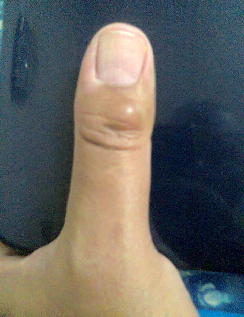
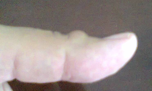
Patients and methods: A healthy man who was born in 1983 consulted us about a small lump on the dorsum of the left thumb. Images are presented, diagnosis is described, and a brief educational review is presented.
Results: The lump was painless and has been noticed more than two years ago. It was cystic in nature and located on the dorsum of the terminal phalanx of the left thumb. It was less than 10 mm in diameter. The diagnosis of digital mucous cyst was made.
Conclusion:
Although consensus has not been confirmed regarding the treatment of digital mucous cyst, small and asymptomatic lesion needs no further treatment or follow-up.
Introduction
Digital mucous cyst is a benign painless skin lesion most commonly observed on the dorsum of the terminal digits, and sometimes on the toes. The lesion generally solitary and less than 10 mm in diameter. It can be opalescent or has the color of the skin. Diagnosis of the condition can be established on clinical basis [1-5].
Patients and methods
A healthy man who was born in 1983 consulted us about a small lump on the dorsum of the left thumb. Images are presented, diagnosis is described, and a brief educational review is presented.
Results
The lump was painless and has been noticed more than two years ago. It was cystic in nature and located on the dorsum of the terminal phalanx of the left thumb. It was less than 10 mm in diameter (Figure-1). The diagnosis of digital mucous cyst was made.
Although consensus has not been confirmed regarding the treatment of digital mucous cyst, the patient in this paper was consulted according to the available evidence suggesting in such small and asymptomatic lesion, no further treatment or follow-up is necessary [4,5].
Discussion
The occurrence of digital mucous cyst was reported as early as the late 1800s, and early 1900s. I was also called myxoid cyst, synovial cyst, and digital ganglion cyst. (Hyde, 1882; Ledderhose, 1893; Ritschel, 1895; Carp and Stout, 1928) [1].
In 1988, Loder et al emphasized the histopathological similarity between digital mucous cysts and dorsal wrist ganglia when examined by light microscopy and scanning electron microscopy. Both lesions had a wall formed by a porous net of collagen fibers, and both had a fibrillated inner surface consisting of randomly arranged collagen fibers, and an intermittent thin membrane. In both lesions, there was no evidence of a cellular lining. Therefore, Loder et al suggested a shared cause for the two lesions [2].
Conclusion
Although consensus has not been confirmed regarding the treatment of digital mucous cyst, small and asymptomatic lesion needs no further treatment or follow-up.
Conflict of interest
None.
References
- Academic Dermatology of Nevada. Digital Mucous Cyst (DMC): Myxoid Cyst. November 25, 2019 [Accessed on the 9th of August, 2022]. https://acadderm.com/digital-mucous-cyst-dmc-myxoid-cyst/
- Loder RT, Robinson JH, Jackson WT, Allen DJ. A surface ultrastructure study of ganglia and digital mucous cysts. J Hand Surg Am 1988 Sep; 13(5):758-62. Doi: 10.1016/s0363-5023(88)80143-6.
- Winterberg DH, Verroen D. Een man met een bobbel op zijn duim [A man with a nodule on his thumb]. Ned Tijdschr Geneeskd 2016;160:A9590. [Article in Dutch].
- Li K, Barankin B. Digital mucous cysts. J Cutan Med Surg 2010 Sep-Oct; 14(5):199-206. Doi: 10.2310/7750.2010.09058.
- Jabbour S, Kechichian E, Haber R, Tomb R, Nasr M. Management of digital mucous cysts: a systematic review and treatment algorithm. Int J Dermatol 2017 Jul; 56(7):701-708. Doi: 10.1111/ijd.13583.


