Current Issue : Article / Volume 3, Issue 1
- Research Article | DOI:
- https://doi.org/10.58489/2836-5038/015
Potency of lysine and tryptophan protect from lysosomal dysfunction and from phenylketonuria, and Mitochondrial disorder mediated by activating lysosomes and OPA1
Biomedical molecular, immunology and Cardiology studies. Toronto Canada and Cairo Egypt
Ashraf M El Tantawi, *
Ashraf M El Tantawi, (2024). Potency of lysine and tryptophan protect from lysosomal dysfunction and from phenylketonuria, and Mitochondrial disorder mediated by activating lysosomes and OPA1. International Journal of Stem cells and Medicine. 3(1). DOI: 10.58489/2836-5038/015
© 2024 Ashraf M El Tantawi, this is an open-access article distributed under the Creative Commons Attribution License, which permits unrestricted use, distribution, and reproduction in any medium, provided the original work is properly cited.
- Received Date: 17-09-2024
- Accepted Date: 30-09-2024
- Published Date: 05-10-2024
Lysine, phenylalanine, tyrosine, phe/hydroxylase, Tyr /hydroxylase, Lysine acetylation, ATPase and GTPase, dopamine, mitochondrial membranes, Nrf2, oxytocin, VEGF-A, angiotensin-2 (Ang2-AT2), phenylketonuria causes,
Abstract
The phenylketonuria due to decreasing in lysine âAAGâ and in Lys/ phosphorylation (which necessary for lysosomal digestive function), and associated with sever decreasing in Trp âTGGâ , followed by mitochondrial dysfunction and lysosomal dysfunction, that followed by sever decreasing in phe /hydroxylase and in Tyr/ hydroxylase , and followed by decreasing in lysine acylation and in SFACs productiin which followed by decreasing in RA productive pathway and associated with increasing in both of TNFa and cholesterol accumulation, which reflect decreasing in estrogen synthesis and in dopamine production, that followed by increasing in the risk of ischemic damage and diabetes.
And we can conclude that both of Lys âAAGâ and Trp âTGGâ are so necessary for activating mitochondrial oxidative functions and necessary for promoting transport system where Mitochondria is so necessary to activate antioxidant function and promoting all of Phe hydroxylase, Tyr hydroxylase, and Lys acylation production which necessary for dopamine and RA synthesis respectively, where the PKU characterized by sever decreasing in antioxidant function, and sever decreasing in both Phe/ hydroxylase & Trp/ hydroxylase production, followed by decreasing in dopamine production and decreasing in retinoic acid production (which due to the reduction in SFACs synthesis).
Retionic acid âRAâ synthesis is activated by lysine acylation and is regulated by lysine acylation mediated by short fatty acid synthesis, which regulate aminoacyl-tRNAs production regulated by mitochondrial enzymes and regulate the mRNA production by E coli (regulated by phenylalanine) that necessary to run antioxidant functions and cellular biosynthesis.
The antihypertensive pathway mechanism is : the Lysine ¬¬> activate ATPase and both lysosomes and Mitochondrial oxidative functions which activate lysine acylation ¬¬> which activate short fatty acid synthesis ¬¬> which stimulate retinoic acid âRAâ synthesis - ¬¬¬> where both Lys and Trp activate mitochondria repair and oxidative functions, which activate, Phe/ hydroxylase, and activate Tyr/ hydroxylase which activate dopamine , and Trp /oxygenases followed by protection against PKU, pulmonary disease ischemic damage and CVD. The activation of antihypertensive pathway associated by activating lysosomes and Mitochondrial oxidative functions, that associated by activating NR4As pathway which responsible for activating GC-beta and both of Oxytocin and Nrf2, followed by Ang2-AT2 and VEGF-A productive functions and heme oxygenase production (notice dopamine regulate heme oxygenase mediated by dopamine beta-hydroxylase which regulated by Mitochondrial oxidative functions) followed by activating anti-inflammatory growth and processes.
Lysine Methylation has important role to enhance and adopt hypertension through activating RA and pervious Antihypertensive pathway mediated by Phe /hydroxylase synthesis and Tyr /hydroxylase production followed by activating both of dopamine and NR4As pathway.
Where dopamine synthesis reflect its productive functions to activate Antioxidant functions that prevent cholesterol accumulation, and in turn increase the estrogen production (which itâs synthesis reflect potential protection from pulmonary disease), and activate the NR4As pathway which regulate all of Oxytocin, Nrf2 production, followed by Ang2-AT2 and VEGF-A production, which followed by activating heme oxygenase and anti-inflammatory growth and processes that prevent LV-hypertrophy, and prevent coronary calcification .
Also, the increasing in Lys AAA with decreasing in Lys AAG and decreasing Trp TGG will cause decreasing in lysosome proper function, and cause Decreasing in GTPase and decreasing in the Mitochondrial oxidative functions, that will cause decreasing in Phe hydroxylase and in Tyr hydroxylase followed by decreasing in lysine acylation, and associated with increasing in cholesterol accumulation, and also associated with decreasing in both of IL17 production and estrogen functional pathway , that will increase the risk of T2D, pulmonary disease, and increase the risk of PKU and CVD.
Both Lys and Trp are necessary to activate transport system across cells membranes that activate antibacterial function and anti-atherosclerosis.
Both Lys and Trp are having the Potency to activate lysosomes and Mitochondrial oxidative functions which activate lipid and protein digestion and enhance Phe hydroxylase and Tyr hydroxylase followed by activating lysine acylation which activate SFACs production and promote the RA synthesis and enhance antioxidant functions followed by dopamine synthesis and protection from ischemic damage and from both of diabetes and PKU
High lights
Lysine “AAG” and Tryptophan “TGG” are so important for activating all of Phe/hydroxylase, Tyr hydroxylase, and Lys acylation for improving own necessary mRNAs production and improving both of protein synthesis and dopamine, where sever decreasing in Lys AAG and Trp TGG cause sever decreasing in Phe /hydroxylase and in Tyr/ hydroxylase.
- the lysosomal damage and mitochondrial dysfunction are associated with phenylketonuria, followed by decreasing in dopamine, and increasing in TNFa which increase the risk of pulmonary disease, CVD, and increasing in Tumor cancer.
- lysosome and Mitochondrial dysfunctions cause increasing in cholesterol accumulation and decreasing in Trp oxygenases production, followed by increasing in TNFa that cause decreasing in short fatty acid production followed by increasing in RA, that cause cause diabetes, coronary artery disease, ischemic damage, and atherogenesis.
_Lys activate lysine acylation mediated lysosomal function and short fatty acid production which necessary for the necessary retinoic acid productive functions.
Methods and results
The Lysosomal dysfunction is associated with increasing in undigested polymers, and decreasing in both E coli and in aminoacyl tRNAs (which regulated by lysine acetylation) productive functions, followed by decreasing in mRNAs (production by E coli) which followed by decreasing in both anti-inflammatory processes and decreasing in heart functions, and followed by increasing in TNFa which activate the increasing in NLRP3 inflammasome functions.
Lysine “AAA, AAG” necessary amino acids for activating lysosomes digestive functions, and necessary to promote both of ATPase and GTPase productive functions, that GTPase necessary to promote mitochondrial repairs and functions which necessary for activating Phe /hydroxylase and Tyr /hydroxylase (where Tyr hydroxylase activate dopamine production), that prevent the cholesterol accumulation and prevent the increasing in TNFa, followed by enhancing the activation of antioxidant function which mediated by activating Phe/ hydroxylase and Tyr/ hydroxylase which regulate dopamine production, then followed by activating NR4As productive pathway which regulate all of GC-beta, B-adrenergic, oxytocin and Nrf2 production .
Decreasing in both of lysine, and Trp, cause mitochondrial dysfunction, and Lysosomal dysfunction followed by increasing in TNFa and appearance of incurable medical cases
Lysosomal dysfunction associated with deficiency in Lysine phosphorylation, followed by decreasing in mitochondrial functions and decreasing in both of Phe/ hydroxylase and Tyr/ hydroxylase which followed by decreasing in E coli functions and decreasing in aminoacyl-tRNAs production, then followed by decreasing in protein biosynthesis mediated by decreasing in mRNAs efficiency (which produced by E coli) , and increasing in both of cholesterol precipitation , and undigested polymers precipitation in blood vessels, then their level in plasma will be elevated , that will be the result of the appearance of pulmonary disease and cardiovascular disease .
The lysosomes dysfunction is due to lysine dysfunction which associated with decreasing in Phe/hydroxylase productive functions which reflect decreasing in mitochondrial oxidative function and in Tyr/ hydroxylase, followed by decreasing in aminoacyl tRNAs production,and decreasing in dopamine biosynthesis , that will lead to abnormal autophagy production and reduction in NR4A pathway that followed by reduction in the antioxidant functions . That, some studies reported: Defective lysosomes lead to abnormal autophagy, activation of inflammation, and reduction in the infection control.[1]
The Lysosomal damage reflect increasing in the undigested polymers and reflect decreasing in GTPase and Mitochondrial dysfunction, that both of cholesterol and undigested polymers will be responsible for activating TNFa production which will activate the NLRP3 inflammasome.
That it has been approved that: the Lysosomal damage has been implicated in activating NLRP3 inflammasome in response to crystals or particulates. [2]
Also, The Lysosomal dysfunction is associated with activating the NLRP3 inflammasome in chronic unpredictable mild stress-induced depressive mice. [3]
So Lysosomal damage reflect increasing in undigested polymers and in cholesterol followed by increasing in TNFa which promote the NLRP3 inflammasomes activities, which will increase the risk of CVD.
The increasing in NLRP3 reflect increasing in TNFa, and reflect severe reduction in both of lysosomal digestive functions and in mitochondrial oxydative function which cause decreasing in both of Phe /hydroxylase and in Tyr /hydroxylase that can cause serious Pathogenic cases eg: diabetes, phenylketonuria, and CVD. That some studies reported that: Activation of Inflammation in Patients with Cardiovascular Disease Are Associated with Higher Phenylalanine to Tyrosine Ratios. [4]
Also, the lysine residues in cathepsin D were so important for phosphorylation as those in procathepsin L. [5]
While, Lysine fatty acylation promotes lysosomal targeting of TNF-α. [6]
So, we can conclude that as lysine fatty acylation biosynthesis activated, as TNFa will be decreased or will be inhibited, that mediated by proper activation to lysosomes and mitochondrial functions.
So lysine is the major amino acid in cathepsin D that is so important for phosphorylation through activating both ATPase and GTPase which necessary for activating mitochondrial repairs and it’s oxydative functions, and activate lysosome digestive functions, therefore prevent the accumulated cholesterol and then prevent the increasing in cholesterol and in both of TNFa and NLRP3, and prevent the aorta and B. vessels calcification.
Where, studies reported that: Lysine functions Prevents Arterial Calcification. [7]
And, the altered CNS excitability due to decreasing in lysine which followed by lysosomal dysfunction and Mitochondrial dysfunction, which followed by increasing in cholesterol and in the undigested polymers which promote the increasing in TNFa production. That studies reported that: the Microglial activation and TNFα production mediate altered CNS excitability following peripheral inflammation. [8]
And so, the lysosomal dysfunction (due to decreasing in lysine phosphorylation) is associated with decreasing in Lys acetylation and decreasing in Phe hydroxylase (which due to decreasing in mitochondria oxidative functions), and then associated with increasing in TNFa, and followed by increasing in the risk of calcification, cardiovascular disease, and pulmonary diseases.
That, Also studies reported that: Lysine suppresses myofibrillar protein degradation by regulating the autophagic-lysosomal system through phosphorylation of Akt in C2C12 cells. [9] note that phosphorylation of Akt is the main for activating lysine Phosphorylation in lysosome that prevent or suppress myofibrillar protein degradation by regulating the lysosomal digestive functions.
Also, lysosome not only is a place for cargo degradation but also plays crucial roles in regulating macrophage polarization. [10] that macrophage polarization mainly regulated by both E coli and mitochondrial functions where their functions mainly controlled by lysosomal function which regulated by the Lys functional activities.
The lysine phosphorylation which activate Phe /hydroxylase and E coli functions is mediated by activating mitochondrial repairs and functions which mainly controlled by GTPase production (that Lys & Trp are having imporoles in activating GTPase production ), where mitochondrial function necessary to activate IL17 production followed by glucocorticoid beta and β-arrestin production and followed by activating both of oxitocin and Nrf2 production (in availability of Leu, Cys, Tyr) to improve myocardial function through adopting glycemic optimal percentages , adopt protein glycation, and form own related macrophages productive functions which adopt anti-inflammatory growth and processes.
Where, studies reported that, L-Lysine is so important to Prevents Arterial Calcification (through activating lysosomal function and activating GTPase which promote mitochondria to prevent cholesterol and undigested polymers accumulation). [11]
Also it has been approved that, Nrf2-mediated anti-inflammatory polarization of macrophages. [12]
Now I would also like to clarify an important fact that: not only lysine important for activating the lysosome and mitochondria, but also the tryptophan, which is very important activator for lysosomal activities and mitochondrial activation through its important ability to produce the GTPase that is used to reactivate the mitochondrial repairs and function.
That it has been approved that: TRP channel 3 (TRPML3), a transient receptor potential cation channel localized to lysosomes. TRPML3 activation initiates lysosome exocytosis, resulting in expulsion of exosome-encased bacteria. [13]
Trp has important role to activate lysosome phosphorylation by activating GTPase production which also has important role in activating mitochondrial repairs and functions, that TRP Channel has important role to Trigger Pathogens Expulsion by activating GTPase production which has the function to activate both lysosome and mitochondria. Also, that previous role of Trp to activate GTPase production can explain its role along the kynurenine (Kyn) pathway to prevents hyperinflammation and induces long-term immune tolerance. [14]
So it’s clear that both of lysine and Tryptophan amino acids are having so important roles in activating both of lysosomes digestive functions and mitochondrial repairs and functions, that both are now responsible for preventing the accumulation of undigested and undigested polymers, then both of Lys and Trp responsible to activate mitochondria which responsible to activate both of IL17 and estrogen, and then are responsible for activating Phe hydroxylase and Tyr hydroxylase production (regulated by mitochondrial oxidative functions) .
And, decreasing in both lysine and Tryptophan are connected to lysosomal damage that can cause decreasing in mitochondrial oxidative function lead to decreasing in both of Phe /hydroxylase and Tyr/ hydroxylase that will cause decreasing in dopamine production and increases in the inflammation that will cause phenylketonuria and and sever decreasing in antioxidant functions.
Also the activation of IL17 is by mitochondrial function will activate GC-beta production and then will activate both of Oxytocin and Nrf2, where Oxytocin alleviates liver fibrosis via hepatic macrophages. [15]
Where, oxytocin stimulates the AMP-activated protein kinase pathway (AMPK) involve the autophagic processes, and may support the renewal of mitochondria. [16]
So, the Treatment with oxytocin can reduces the accumulation of proinflammatory cytokines and reduces immune cell infiltration. That Oxytocin stimulates differentiation stem cells to cardiomyocyte lineages as well as generation of endothelial and smooth muscle cells, promoting angiogenesis. [17]
Also it has been approved that: Tryptophan metabolism important for treatment patients suffering from CVD. [18]
So CVD is due to sever decreasing in lysosomes in mitigating region and functions that can be treated by tryptophan and lysine to which can activate both previous intracellular system to recover CVD.
Notice that High tryptophan diet reduces extracellular dopamine, and can affect the dopamine levels and functions which will be discussed later.
Lysosomal dysfunction is associated with increasing in TNFa and decreasing in VEGF-A
Firstly, the Lysosomal dysfunction induce apoptosis in chondrocytes through BAX-mediated mitochondrial damage. [19]
And the mitochondrial dysfunction promise the increasing and accumulation in cholesterol and pro-inflammation, that it has been approved that Chronic inflammation during IBD may be mediated by NLRP3 inflammasome activation. [20]
And, free Cholesterol-loaded Macrophages Are an Abundant Source of Tumor Necrosis Factor-α and Interleukin-6. [21]
And now, the Increasing in TNFa is related to increasing in cholesterol and decreasing in both of mitochondria and lysosomes digestive function, will be formed by decreasing in myocardial function. Where, TNF-alpha downregulates expression VEGF receptors in human endothelial cells [22]
The Lysosomal dysfunction is associated with decreasing in mitochondrial function followed by increasing in both of cholesterol and TNFa production , where TNFa activate NLRP3 inflammasome pathway functions followed by enhancement the cardiovascular pathologies development .
That studies reported that: the Lysosomal dysfunction is associated with NLRP3 inflammasome activation. [23]
And the TNf-a regulates transcription of NLRP3 inflammasome components and inflammatory molecules. [24]
Also, the increasing in lysosomal dysfunction reflect decreasing in lysine followed by decreasing in GTPase and decreasing in mitochondrial function then followed by increasing in undigested polymers and increasing in TNF-α which enhanced the development of a number of cardiovascular diseases. [25]
Also, Patients with acute myocardial infarction had statistically significant increased serum levels of PTEN & TNF-α gene activity. [26]
So, it’s clear that Patients with acute myocardial infarction are having severe Decreasing in both lysosomal and mitochondrial functions followed by increasing in the accumulated cholesterol, undigested polymers, and TNFa functional activities, which originally caused by sever decreasing in both of lysine phosphorylation and Tryptophan functions, which followed by decreasing in dopamine synthesis.
That both DPM and DBM are useful in acute cardiogenic circulatory collapse. [27]
That previous study indicated that CVD characterized by severe reduction in dopamine, that’s why dopamine is useful for treating patients with CVD.
And, the Myocardial Infarction is a Consequence of Mitochondrial Dysfunction. [28]
And, In classical PKU, the serotonin and dopamine biosynthesis is inhibited. [29]
So it’s clear that both of CVD and PKU are characterized by severe reduction in dopamine, that means those two diseases are connected together in lysosomal and mitochondrial dysfunction, followed by increasing in the accumulated cholesterol, undigested polymers, and TNFa functional activities, which originally caused by sever decreasing in both of lysine phosphorylation and Tryptophan functions.
Also, the Tumor Necrosis Factor-α (TNFa) promotes and exacerbates calcification in heart valve myofibroblast populations. [30]
Also, studies approved that the TNF-α antagonism is unlikely to be a beneficial therapeutic strategy in patients with acute myocardial infarction. [31]
Also, the lysosomal dysfunction can Increase the concentrations of TNF-α which are found in acute and chronic inflammatory conditions. [32]
Now, normally and positively the Genetically-predicted TNF levels are associated with coronary artery disease and inversely associated with overall cancer. [33]
While, Lysine Prevents Arterial Calcification in adenine-induced uremic rats. [34] And Nuclear ATR lysine-tyrosylation protects against heart failure. [35]
Where, cerebral ischemia due to extensive calcified vasculopathy, disruption of the basal ganglia-thalamo-cortical circuit, and nigrostriatal dopaminergic dysfunction are plausible pathogenic mechanisms. [36]
So, lysine and Tryptophan are playing important role in Protecting from cerebral ischemia, Heart Failure, and also Prevents Arterial Calcification by enhancing lysosomal Functions and mitochondrial repairs and functions followed by enhancing estrogen production which prevent the accumulated cholesterol and un-digestive polymers. [37]
Where, VEGF production regulated by angiotensin-2 which controlled by mitochondrial function, which regulated by intracellular ATPase and GTPase
That studies approved that, mitochondrial function control delivery of nutrients and oxygen to tissues through the extension of blood vessels by angiogenesis. [38]
So, decreasing in lysosomal function followed by reduction in mitochondrial function will increase all of cholesterol TNFa production and reduce angiotensin-2 and VEGF-A productive functions that can be the result of of increasing the risk of cerebral ischemia, CVD, and Arterial Calcification, followed by reduction in neuroprotective effects on hypoxic motor neuro, and decreasing in the protection against memory impairment “figure 1 “ “which Explains, describes and summarizes the basics of the importance of pegsin and trytophan in this work”.
Where, VEGF-A also has a neuroprotective effect on hypoxic motor neurons, and is a modifier of ALS (amyotrophic lateral sclerosis). [39]
Also, it is important to note that: VEGF A in serum protects against memory impairment in APP/PS1 transgenic mice by blocking neutrophil infiltration. [40]
While, producing IL17 activated by mitochondrial function where IL17 is important activator to Glucocorticoids-beta synthesis via NR4As pathway (and prevent organs failure cholesterol accumulation), followed by activating B-arrestins and both of B-adrenergic and Nrf2 synthesis followed by activating angiotensin-2 (Ang2-AT2) and VEGF-A productive functions. [41]
Also, both of Lys and Glu (Lys AAG is reversed copy of Glu GAA) are having important role in stabilizing leucine CTT functional pathway and are important for activating mitochondrial function which regulate il 17 production which activate NR4As pathway started by activating GC-beta, followed by activating angiotensin-2 and VEGF-A production, which reflect reduction in TNFa and cholesterol.
So, Lysosomal dysfunction is associated with decreasing in mitochondria function and decreasing in VEGF-A followed by increasing in TNFa, where TNFa activate NLRP3 inflammasome pathway functions followed by activating the cardiovascular diseases, myocardial infarction as described in “Figure 1”, (figure 1is explanation of main aspect of the importance of lysine & Trp in lysosome & Mitochondrial oxidative functional pathways).
Both lysine and phenylalanine important for treating pulmonary disease
Firstly, pulmonary diseases characterized by mitochondrial dysfunction followed by increasing in cholesterol accumulation, and followed by defect in estrogen biosynthesis (where cholesterol is the substrate for estrogen synthesis regulated by synthase enzyme), followed by decreasing in IL17 and GC-beta production and decreasing in NR4As pathway, that it has been approved that
Estrogen Inhibition Reverses Pulmonary Arterial Hypertension and Associated with Metabolic Defects. [42]
Decreasing in Estrogen reflect increasing in cholesterol accumulation due to decreasing in OPA1 repair and functions, where mitochondrial repairs and functions provided by GTPase which produced by Trp “TGG “and Lys “AAG ” , that conclude :the estrogen Inhibition Reflect Inhibition in mitochondrial functions that reflect sever decreasing in Lys and in Trp, that followed by Inhibition in NR4As pathway and Inhibition in both of angiotensin-2 and VEGF-A production, followed by increasing in TNFa, and increasing in the risk of Pulmonary Arterial Hypertension, and arterial calcification.
Also, we can conclude that lysosomal dysfunction and Mitochondrial dysfunction is the main for plumbing disease
Also, mitochondrial dysfunction is a systemic phenomenon during chronic obstructive pulmonary disease COPD [43]
Also, studies reported that Mitochondrial Dysfunction as a Pathogenic Mediator of Chronic Obstructive Pulmonary Disease and Idiopathic Pulmonary Fibrosis. [44]
And, the absence of Prolyl-4 Hydroxylase 2 (PHD2) indicate absence of Α-Ketoglutaric acid which is a short-chain fatty acid, that reflect sever decreasing in mitochondrial function and sever decreasing in both Lys and Trp that will be the result of pulmonary disease survival and can cause another Incurable diseases.
That it has been approved: the Prolyl-4 Hydroxylase 2 (PHD2) Deficiency in Endothelial Cells Induces Obliterative Vascular Remodeling and Severe Pulmonary Arterial Hypertension. [45]
Also, studies indicated that: GTP-bound Rab7 promotes mitochondria–lysosome contact site formation and tethering, while mitochondrial TBC1D15 (Rab7-GAP) recruited to mitochondria via Fis1 drives lysosomal Rab7 GTP hydrolysis at mitochondria–lysosome contact sites. [46]
So, pulmonary disease characterized by Inhibition Ras proteins which control mitochondrial function, followed by Inhibition in GTP bound Rab7, and followed by mitochondrial dysfunction and accumulation in cholesterol and increasing in TNFa functions.
Where, Ras proteins control mitochondrial biogenesis and function. [47]
Also, Lysine acylation synthesis reflect Activation to Mitochondrial Aconitase in the Heart [48], mediated by activating ATPase and GTPase production, and followed by preventing the accumulation of cholesterol and protect from pulmonary disease.
Also, alterations in the expression of Lysine Methyltransferase have been linked to the genesis and progress of several diseases, including cancer, neurological disorders, and growing defects, [49]
So, the availability of lysine is necessary for lysine Acetylation synthesis mediated by Activating Mitochondrial Aconitase in the Heart through activating GTP-bound Rab7 which promotes mitochondrial and lysosome functions followed by preventing the cholesterol accumulation, and followed by activating Phe hydroxylase production which controls the phenylalanine metabolic functions, and followed by preventing aorta calcification.
Also, the absence of lysine will inhibit lysine fatty acylation followed by Inhibition to adipocyte B-adrenergic function which will be followed by decreasing in fatty metabolic process lead to cholesterol accumulation. That the presence of lysine is necessary to activate the lysine fatty acylation of an anchoring protein mediates adipocyte adrenergic functions (which has the antioxidant function). [50]
Also in the other hand, TBX5 is required for patterning of the cardiac system and maintenance of cardiomyocyte functions, where lysine is required for acetylation which required for activating TBX5 for Regulating heart development and function ,“figures1&3”. [51]
So, both of Lysine and Trp have so important roles in activating lysosomes and mitochondrial function which followed by activating Lysine fatty acylation which required for activating TBX5 function and antioxidant mediated by Phe /hydroxylase and Tyr/ hydroxylase productions which considered as stable mRNAs produced by E coli for modulation heart and brain functions, and necessary for controlling phenylalanine metabolic pathway.
I would like to give a little note that , arginine has important role in activating tRNAsheart and accelerate transport system pathway , that Arg is required to be included in mRNAs which produced by E coli to accelerate immune functional pathway that accelerate antioxidanty and transport systematic pathway (which mainly regulated and promoted by GTPase production ) function and prevent calcification and the accumulation of inflammation which can be the main for lysosomal and Mitochondrial disorders.
That it has been approved that: l-Arginine prevents the over-expression of alkaline phosphatase (ALP, p less than 0.001) and reduces matrix calcification (p less than 0.05) in VICs treated with LPS. [52]
And, Homo-arginine Supplementation Prevents Left Ventricular Dilatation and Preserves Systolic Function in a Model of Coronary Artery Disease. [53]
And, decreasing in both of lysine and Trp will decrease mitochondrial oxidative function followed by decreasing in Lys acylation which followed by decreasing in TBX5 functional activities and decreasing in heart function and development , and also the decreasing in lysosome and Mitochondrial functions will be followed by increasing in inflammation and in cholesterol accumulation, and will cause decreasing in phe/hydroxylase production which cause increasing in the risk of both of PKU and the cardiovascular disease that it has been approved that : the cardiovascular phenotype of adult PKU patients is characterized by high levels of inflammatory and oxidative stress markers, endothelial dysfunction and vascular stiffness “figure 2 “. [54]
Also, it has been approved that the abnormalities in patients with phenylketonuria consisted of widening and cupping of the metaphysis, an intact zone of provisional calcification.[55]
And, the patient with malignant phenylketonuria (PKU) who underwent both CT and MR Imaging is reported, that CT demonstrated the characteristic calcifications of the basal ganglia. [56]
Also, decreasing in pyrimidine kinases (pyrimidine synthesis regulated by mitochondrial synthetase) reflect mitochondrial dysfunction, followed by increasing in cholesterol accumulation and decreasing in phenylalanine hydroxylase, which reflect reduction in antioxidant function, which reflect reduction in estrogen synthesis and decreasing in dopamine synthesis, which followed by aorta classification and pulmonary disease.
Where, studies reported that: Deficiency in pyrimidine kinases cause cholesterol accumulation and increase the risk of coronary artery disease. [57]
So, decreasing in Phe /hydroxylase reflect decreasing in both of lysine and Tryptophan followed by decreasing in lysosomes function which followed by decreasing in mitochondrial oxidative function that will be result in decreasing in both of Phe hydroxylase synthesis and in Lys acylation (which required for TBX5 function), and then the decreasing in lysosome and in mitochondria function will cause increasing in cholesterol and in TNFa which will be followed by increasing phenylalanine accumulation which will be followed by increasing in calcification and pulmonary disease “Figure 1 and 2 “.
Also, phenylalanine and Phe/hydroxylase controlled by lysine functions through translation and by activating mitochondrial functions, that will be the reason of protecting Against monocrotaline-induced pulmonary vascular remodeling and lung inflammation. Where studies indicated that 4-Chloro-DL-phenylalanine protects against monocrotaline‑induced pulmonary vascular remodeling and lung inflammation. [58]
So again, the lysine is basic controller to Phenylalanine and phe/hydroxylase
Functions by translation process Lys “AAA, AAG “¬¬>Phe “TTT, TTC” where, both of lysine and Tryptophan TGG are the basic activator to mitochondrial repairs and oxidative functions which activate all of Phe /hydroxylase, Tyr hydroxylase followed by dopamine productive functions.
Lysosomal dysfunction cause decreasing in mitochondria and in both of lysine Methylation and Phe /hydroxylase, where Lys and Trp required for alveoli and protect from pulmonary, and phenylketonuria
As we mentioned before lysine phosphorylation is the main activator for lysosomal function, where other studies approved that: the Loss of laat-1 transporter caused accumulation of lysine and arginine in enlarged, degradation-defective lysosomes. [59]
And, lysine residues are required for phosphorylation of procathepsin L and are a common feature of the site on many lysosomal proteins. [60]
That, as lysine decreased as so many of lysosomal protein will be decreased and cause lysosome damage.
Also, both of Lys “AAG” and Trp “TGG “are so necessary for activating mitochondrial oxidative functions which is so necessary to activate antioxidant function where PKU characterized by sever decreasing in antioxidant function. That, oxidative stress is involved in the pathophysiology of the tissue damage found in PKU. [61]
Also, decreasing in Phe /hydroxylase reflect decreasing in mitochondrial oxidative functions which demonstrate the energy deficit and oxidative stress which characterized PKU pathophysiology. [ 62]
_And the Mitochondrial dysfunction cause cellular injury and declined antioxidant capacity. [63]
_where, the mitochondrial redox homeostasis is a potential target for disease treatment,
That disruption of mitochondrial redox homeostasis in muscle resulted in energy defect and exercise intolerance. [64]
_And, Mitochondrial necessary to activate antioxidant which the step toward disease treatment. [65]
Also, Tryptophan “TGG“ has the role of reducing the mRNA levels of proinflammatory cytokine genes and enhance mitochondrial function by increasing the mRNA levels of mitochondrial transcription.[66]
_While Dietary lysine levels modulate the lipid metabolism, mitochondrial biogenesis and immune response.[67]
And, also, it’s very important to note that Lysine Acetylation has the roles of Activating Mitochondrial Aconitase in the Heart [ 68], which mediated by short fatty acid production which activate retinoic acid function that adopt heart function
That Short-chain fatty acids activate acetyltransferase p300. [69]
Where, and lysine acyltransferases regulate protein synthesis, and are direct sensors and mediators of the cellular metabolic state.[70]
And also, the short fatty acids (SCFAs) promote retinoic acid synthesis and functions, that SCFAs crosstalk with RARα in dendritic cells as a critical modulator that plays a core role in promoting Th2 cells differentiation at a state of modified/disturbed microbiome, “figure 1”. [71]
So, we can conclude that both of Lys and Trp are so necessary for activating mitochondrial oxidative functions which is so necessary to activate antioxidant function which promote Phe/ hydroxylase & Tyr/ hydroxylase production, where the PKU characterized by sever decreasing in antioxidant function and sever decreasing in Phe hydroxylase production.
Also Lys acylation has the importance role in activating short fatty acid chains “SFACs“ and acetyltransferase production which mediated the importance of proper metabolic pathways, where SFACs have important roles for activating retinoic acid functional pathway which has important role to activate antioxidant , that’s why PKU characterized by reduction in Phe/ hydroxylase, (and reduction in Tyr/ hydroxylase due to reduction in mitochondrial function ) reduction in acetyl transfers, and then reduction in SFACs production followed by reduction in both of retinoic acid function and reduction in dopamine productive functions.
Therefore, the lysine has an intense role in activating immune functions and anti-inflammatory processes, that the necessity of the Regulation of lysine methylation emerged as a critical regulator of neurological function and disease. [72]
Again the meaning of the necessity of Lys methylation to be produced in vivo is reflecting the proper availability of lysosomal and mitochondrial function that reflect Phe/ hydroxylase productive pathway and Tyr/ hydroxylase pathway, and also reflecting the short fatty acid production which necessary for activating retinoic acid synthesis which necessary for activating RORs functional pathway and activating antioxidant function , which followed by activating NR4As pathway that enhance full controlling the antioxidant functions through producing oxytocin and Nrf2, and full angiotensin-2 and VEGF-A functional pathway which activate anti-inflammatory growth and processes to protect from many viral toxins and protect from many critical diseases including mitochondrial disorders and inflammatory disorders.
The Mitochondrial dysfunction and lysosomal dysfunction are reflecting reduction in lysine acylation and in Trp, which reflect decreasing in short fatty acids “SFACs” production which followed by reduction in RA (which regulated by Lys acylation & SFACs ), and followed by increasing in cholesterol which activate increasing in TNFa that play important roles in inflammatory diseases, which characterized by swelling due to absence of lysosomal and Mitochondrial functions, where Some oncologists can be confused about swelling in tissue that can consider it as tumor cancer without understanding the basic of swelling.
Also, dopamine (regulated by Trp) may have beneficial effects on the respiratory system by decreasing oedema formation and improving respiratory muscle function [73]
That Dopamine Reverses Lung Function Deterioration After Cardiopulmonary Bypass Without Affecting Gas Exchange. [74]
So, The decreasing in both of lysine “AAG” and Tryptophan ”TGG” cause decreasing in lysosomal function and in mitochondrial oxidative function, that will cause accumulation in the undigested polymers which cause lysosomal enlargement, then followed by decreasing in lysine acylation and in aminoacyl tRNAs, that will cause decreasing in Phe/ hydroxylase and decreasing in Tyr/ hydroxylase, followed by decreasing in E coli and decreasing in dopamine productive functions, and followed by decreasing oedema formation and improving respiratory muscle functions ”figure 1& 2”.
Lysine and Tryptophan as discussed before are so important for activating mitochondrial oxidative function which necessary for both of pyrimidine kinases, and estrogen synthesis (which required for pulmonary alveolar functions).
That it has been approved that, estrogen receptors, are required for the formation of a full complement of alveoli in female mice. [75]
Now, we can understand why both of lysine and Tryptophan are so important for treating pulmonary disease, and increasing in Left vertical size (due to increasing in cholesterol and in TNFa, and decreasing in GTPase and in estrogen due to decreasing in mitochondrial function ), where Lys phosphorylation roles and Tryptophan are important for activating mitochondrial functions, mediated by activating both lysosomes and E coli functions, and followed by activating Lys acylation and Phe /hydroxylase productions .
Also, the Aztreonam lysine “AZLI” (an inhaled lysine salt formulation) is effective, safe and well tolerated in the treatment of acute pulmonary exacerbations of CF. Superior improvements in lung function and quality of life suggest AZLI may represent a new treatment approach for acute pulmonary exacerbation. [76]
And for sure the decreasing in mitochondria function will be followed by decreasing in phe /hydroxylase and in Tyr hydroxylase, then will be followed by accumulated Phenylalanine , and reduction in dopamine biosynthesis, which followed by increasing in oedema formation and decreasing in respiratory muscle functions “figure 2”.
Also, the Inhibition in dopamine (due to Inhibition in both of Lys /hydroxylase and Phe /hydroxylase) is Associated with phenylketonuria and connected to lysosomal dysfunction.
Where, Phenylketonuria (PKU) is an autosomal recessive disorder caused by reduction in phenylalanine hydroxylase, which required for tyrosine hydroxylase synthesis, which is essential for dopamine production. [77]
Also, adult Patients Suffering from Phenylketonuria are suffering from reduction in Cerebral Fluoro-l-Dopamine Uptake. [78]
Where , the accumulation of phenylalanine in the blood of patients suffering from phenylketonuria, that are suffering from deficiency in both of lysine hydroxylase and Phenylalanine hydroxylase will be the cause of cause accumulation in phenylalanine followed by reduction in Tyr/ hydroxylase and reflect reduction in all of GTPase and mitochondrial function, that will cause reduction in dopamine synthesis (and reduction in antioxidant and in the formation of oedema formation and associated with reduction in respiratory muscle function ), ” figure : 1& 2”. [79]
So, sever decreasing in Lys and Trp will cause the lysosomal damage and mitochondrial dysfunction followed by the occurrence of such phenylketonuria (or the appearance of phenylketonuria)) which Associated with reduction in Phe/ hydroxylase and reduction in Tyr /hydroxylase (which considered a sign of phenylketonuria) and associated with decreasing in dopamine, followed by increasing in the risk of pulmonary disease, CVD.
Again, not only lysine are important to regulate mitochondrial oxidative functions, but also tryptophan which GTPase that promote mitochondrial repairs and functions and also is important to promote dopamine production which is reported that Trp is necessary to regulates DAergic neurogenesis and Mitochondrial oxidative functions. [80]
And, both of lysine and Tryptophan are having important roles in activating mitochondrial oxidative function that enhance the Phe/hydroxylase and Tyr hydroxylase synthesis and prevent the accumulated phenylalanine and then control it’s improved metabolic pathway which necessary for protecting both of heart and brain function, and also are important for promoting IFNs production which will be discussed later.
Also, lysine acylation necessary for activating E coli, and allows the actual effects of lysine acetylation for protein productive functions. [81]
Also, as lysine necessary for activating lysosomal function, as dopamine also is very important for promoting lysosomal Functions and is strongly connected to lysine functions for promoting both of lysosomes and mitochondria function.
That it has been approved that Inhibition of the dopamine transporter promotes lysosome biogenesis and ameliorates Alzheimer's disease-. [82] And so, we can conclude that: both lysine phosphorylation and Tryptophan are connected together in their functions for activating lysosomal function and mitochondrial function, followed by activating Phe/ hydroxylase and Tyr/ hydroxylase production, then followed by their roles in activating E coli functions, and the necessity of Lys acylation in activating retinoic acid synthesis mediated by activating SFACs production.
Lysine, phenylalanine, and tryptophan, promote dopamine synthesis and Mitochondrial functions that protect from alzheimers disease
Firstly, as we discussed previously that both lysine and Tryptophan are having their strong roles in regulating all of lysosomes, mitochondria, and E coli functions, as sever decreasing in those amino acids can lead to sever decreasing in lysosomes and in E coli that will be the result of decreasing in heart function that lead to ji
cardiac impairment.
That, patients suffer from a lack of aminoacyl tRNA productions and they suffer from lysosome and E coli dysfunctions. [83] Where previous study indicated decreasing in Lys will cause reduction in Lys acylation which will be followed by reduction in aminoacyl tRNAs synthesis and mainly cause reduction in lysosomal function which will be followed by increasing in accumulated undigested polymers and cholesterol and reduction in retinoic acids synthesis which regulated by SFACs which it’s production regulated by Lys acylation function.
And, the modulation of activating mitochondria and both of lysosomes and E coli can be a potential therapeutic strategy for age-associated cardiac impairment. [84]
Also, Nuclear ATR lysine-tyrosylation protects against heart failure by activating DNA damage response. [85]
And other studies approved that l-Lysine is an essential amino acid important for maintaining human health. [86]
Now in the availability of proper mitochondrial function: the controlled increasing in Phenylalanine synthesis will stimulate the increasing in immune antioxidant through Phe/ hydroxylase production (regulated by mitochondrial oxidative function), which increase the E coli functions and activate Tyr hydroxylase which will activate dopamine production (where dopamine considered as strong activator to antioxidant functional pathway). [87]
And, both of Phenylalanine (Phe) and tyrosine constitute the two initial steps in dopamine biosynthesis “figure 1”. [88]
Notice that, both of tryptophan and Tyr/ Hydroxylase Regulate Dopamine Synthesis, where studies has been approved that: the Tyrosine hydroxylase (TH) catalyzes the rate-limiting step in the biosynthesis of dopamine (DA) and other catecholamines, and its dysfunction leads to DA deficiency and parkinsonisms. [89, 90]
Also, the parkinson Diseases characterized by depletion of dopamine (which regulated by tryptophan, and by both of Phe/ hydroxylase and Tyr/ hydroxylase synthesis). [91]
Also, The Dopamine (productive pathway) has the role of increasing the force of contraction, and increase the elevation of the beating rate, and the constriction of the coronary arteries. [92]
Whereas mentioned previously both of lysine and Tryptophan activate mitochondrial function that promote both of Phe/ hydroxylase and Tyr/ hydroxylase production which activate dopamine production “figure 1&2”, while mitochondrial synthase promote IL17 production which activate GC-beta, followed by activating B-adrenergic and both of Oxytocin and Nrf2 production which considered as strong antioxidant.
And now we can understand that the mechanism of dopamine is necessary for promoting anti-hypertensive pathway by lowering blood pressure. And the decreasing in Phe / hydroxylase and in Tyr /hydroxylase will be followed by decreasing in dopamine and decreasing in antihypertensive pathway, “Figure 4&5”. Where studies approved that: Dopamine considered as promoting antihypertensive pathway, by lowering blood pressure. [93]
Also, Dopamine b: is considered as Important Antihypertensive Counterbalance Against Hypertensive Factors. [94]
Where, the antihypertensive pathway mechanism is : the tryptophan and Lysine ¬¬> activate mitochondrial repairs and functions ¬¬> will activate methylation ¬> which will activate Phe/ hydroxylase ¬¬> followed by activating Tyr/ hydroxylase ¬> followed by activating dopamine ¬>where dopamine synthesis in vivo which necessary to lowering “adopt” blood pressure “figure 1& 4” , also reflect the activation to NR4As pathway mediated by activating the IL17 by mitochondrial synthase function ¬> which activate GC-beta production ¬> followed by activating both of Oxytocin and Nrf2 ¬> Ang2-AT2 and VEGF-A productive functions ¬>heme oxygenase ¬>anti-inflammatory growth and processes.
Also, Tyr / hydroxylase are found in heart tissue that indicated the necessity of dopamine in adopting heart functions directly and indirectly, that studies reported that: the activity of the enzyme tyrosine hydroxylase showed that was present in both hearts and in tissue with total sympathetic denervation. [95]
So, dopamine (regulated by tryptophan, Phe/ hydroxylase, and Tyr/ hydroxylase) are having the role of protecting heart function and brain function through activating antioxidant function and enhance mitochondrial function, followed by lowering “adopt” blood pressure.
Also, it is reported that: L-lysine influences the selective brain activity in dependence on the biological significance of pain-induced behavior. [96]
So it is clear that lysine and Tryptophan are so important to improve lysosomal function followed by enhancing the activation of mitochondrial proper functions followed by activating both of dopamine and antioxidant (by activating antihypertensive pathway) mediated by Improving both of Phe/hydroxylase and Tyr hydroxylase y “Figure 1&4” , followed by activating NR4As pathway, which mediated by activating IL17, that promote GC-beta and both of oxytocin and Nrf2 production which necessary for adopting antioxidant functions.
That, DA has direct autocrine effects on beta cells, and indirect paracrine effects through delta cells. [97]
Where, deficiency in Phe/ hydroxylase followed by decreasing in both of antihypertensive pathway, and decreasing in Tyr/ hydroxylase will cause decreasing in all of lysosomal function and diseasing in mitochondrial function, and then decreasing in dopamine, which followed by the increasing in Blood pressure and decreasing in antioxidant function , that will be the result of causing PKU symptoms which is characterized by an accumulation of traditional cardiovascular risk , high levels of inflammatory and oxidative stress markers, endothelial dysfunction and vascular stiffness . [98]
Decreasing in Retinoic acid related orphan “RORs” is fully connected to lysosomal dysfunction and mitochondrial dysfunction that cause increasing in both of TNFa and cholesterol
There are strong cooperation between Lys, Trp, and RORs productive functions in activating immune functionality , that both Lys and Trp are very necessary for activating both of lysosomes and functions and mitochondrial functions which responsible for promoting RORs productive pathway that their activities reflect Inhibition to cholesterol accumulation and activate both of IL17 and estrogen productions, and also their activities consequently inhibits TNFa production (through activating lysosome and Mitochondrial functions).
And, I’ve mentioned previously that absence of lysine will inhibit lysosomal function and will inhibit lysine fatty acylation synthesis which necessary for SFACs production which by itself is necessary for Regulating retinoic acid productive , and absence of lysine will be followed by Inhibition to adipocyte B-adrenergic function, which followed by decreasing in fatty metabolic process and increasing in both of undigested polymers and cholesterol accumulation, then cause decreasing in Retinoic acid functional pathway . That retonic acid functional pathway is fully connected with and regulated by lysosomal function and by mitochondrial function, that Mitochondrial functions is regulated by both lysine and Tryptophan functional pathways, where retinoic acid functions promote the decreasing in TNFa functions.
That studies reported that: Retinoic acid receptor-related orphan receptor A (RORA) inhibits the transcription and expression of TNF,” Figure 1”. [99]
So, it’s very clear that Lys and Lys acylation are playing so important role in decreasing TNFa through the role of Lys acylation in activating SFACs production which necessary for activating retinoic acid productive functions.
While, the short fatty acids “SFACs” production which promoted by lysosomal digestive functions and by mitochondrial oxidative function (and activate Lys acylation which important for SFACs production) are very important for retonic acid synthesis, where in case of lysosomal dysfunction and Mitochondrial dysfunction will cause reduction in lipid digestion and sever reduction in short chain fatty acids “SFAC”. That it has been approved that “SCFAs” affect DCs by facilitating retinoic acid synthesis “Figure 1”. [100]
Retinoic acid-related orphan receptors RORs (regulated by mitochondrial function) play a regulatory role in lipid/glucose homeostasis and various immune functions.
That decreasing in Trp “TGG” and in Lys “AAG” will cause reduction in both of lysosomes and mitochondrial functions, and will be associated with increasing in cholesterol and in undigested polymers which promote the increasing in TNFa productive functions.
There are strong cooperation between Lys, Trp, and RORs productive functions in activating immune functionality, and in protecting from both of diabetes and calcification, that both of lysine “AAG” and Tryptophan” TGG “are having important role in the decreasing all of cholesterol, TNFa, and Calcifications, followed by protection from tumor cancer.
The Retinoic acid is necessary for Retinoic Acid Receptor-Related Orphan Receptors for running several cellular metabolic functions, that reduces apoptosis and oxidative stress, evaluate cardiac development, enhances the repair of infarcted myocardium, and the all-trans retinoic acid “ATRA” is having important role in dissolving Aorta calcification by promoting fission events throughout accelerating lysine and Trp functions for activating mitochondrial repairs and functions
Where studies approved that: Retinoic acid (RA) established several functions during cardiac development, including actions in the fetal epicardium required for myocardial growth, (also, retinoic acid reduce oxidative stress),” figure 1”. [101]
And the activated acyclic retinoic acid pathway can reflect activated ribosomes and mitochondrial function for activating lysine acylation which necessary for activating SFACs production followed by activating Phe/ hydroxylase and Tyr/ hydroxylase production and IL 17 production from acting on IL2&6 (which means previous processes control the Dendritic cells function), followed by activating GCs-beta and both of oxytocin and Nrf2 which followed by activating angiotensin2 and VEGFa functional pathway.
Where studies approved that, acyclic retinoic acid pathway can specifically block calcification in a vascular cell.[102]
I would like to mention that: it’s clear the high level of endothelial dysfunction is the sign of mitochondrial dysfunctions, and reflect sever decreasing in lysine and Tryptophan and reflect increasing in cholesterol and decreasing in TNFa followed by un controlling to Dendritic Cells functions and then followed by increasing in vascular stiffness.
Also notice that, as both of lysosomes and mitochondrial dysfunction increased as the function of RORs will be decreased and will be associated with decreasing in antioxidant function and decreasing in lipid metabolic pathway that will be associated with decreasing in SFACs production , followed by increasing in TNFa and increasing in the risk of high blood pressure and increasing in the risk of heart failure, that studies approved that : the proteomic analyses of human and guinea pig heart failure (HF) were consistent with a decline in resident cardiac all-trans retinoic acid ”ATRA” “Figure 1”. [103]
Also, the reduction In ROR pathway reflect Diabetic cardiomyopathy. [104]
So , Reduction in RORs pathway reflect decreasing iny retinoic acid synthesis and then decreasing in Lys-acylation production which followed by decreasing in SFACs production and reduction in both of lysosomes and mitochondrial functions that reflect defect in both of Lys “AAG” and Trp “TGG” availability, followed by reduction in GTPase production and then reflect reduction in RORs subunits production (which regulated by OPA1 function ), then followed by reduction in the estrogen production and increasing in the accumulated androgen , and Associated with accumulation in both of cholesterol and undigested polymers which promote the increasing in TNFa, which followed by decreasing in dopamine production and increasing in Blood pressure followed by increasing in calcifications, and cause Diabetic cardiomyopathy.
Where, studies approved that The Retinoic acid promote the increasing in the mRNA levels and protein synthesis of matrix Gla protein, and calcification inhibitory molecule, in human coronary artery and aortic valve cells. [105]
The sever decreasing in Lys “AAG” and in Trp “TGG” promise decreasing in lysosomal Functions which play important role in atherogenesis, CVD and in coronary calcifications
The lysosomes are
Membrane-bound organelles with roles in processes of degrading and recycling cellular waste, cellular signalling and energy metabolism. Sever decreasing in lysine and in tryptophan (decreasing in dopamine) cause lysosomal storage disorders, survival of tumor cancer, autoimmune disorders, blood disorders, Atherosclerosis, and neurodegenerative diseases.
The lysosomal dysfunction promise the accumulated phenylalanine, where the absence of lys AAG reflect decreasing in the translations and stability of the normal Phe functions that will cause Phe accumulation.
Also, sever decreasing in lysosomal functions cause decreasing in mitochondrial function that cause decreasing in lysine acylation and in SFACs production and followed by decreasing in retinoic acid synthesis and reduction in antioxidant pathway which followed by increasing in inflammation, and in cholesterol deposit in arteries walls that cause calcification.
As I discussed previously, both of lysine and Tryptophan are necessary for activating mitochondrial oxidative functions and both necessary for reducing inflammation, and cholesterol Mitochondrial disorder through increasing fat digestion and producing SFACs which activate RA synthesis and activate both of RORs pathway and antioxidant function , followed by decreasing or inhibiting the increasing in TNf-a, but now not only lysine phosphorylation necessary for lysosomal Functions but also tryptophan is so necessary for protecting and purifying lysosomal Functions and contribute in the SFACs production and in RA productive functions which begin by activating mitochondrial functions and increasing the cellular transport system functions, . And also the necessity of Trp can be started by activating mitochondrial oxidative functions which activate both Phe hydroxylase and Tyr hydroxylase, and activate tryptophan-2,3-dioxygenase which necessary for protecting heart from ischemic damage.
Where, studies approved that: Tryptophan
“TRP” Channel Senses Lysosome Neutralization by Pathogens to Trigger Their Expulsion. [106]
And, as we mentioned previously that Lys which activate lysosomal function is so necessary for translating and stabilizing Phe functions, that studies reported that: Involvement of lysosomes in substrate stabilization of tryptophan-2,3-dioxygenase. [107]
And other studies reported that : The tryptophan oxygenase are so necessary for protecting heart from ischemic damage, “figure 1”. [108]. So, lys is so necessary to promote and stabilize Phe functional stability which promote tryptophan dioxygenase which protect heart from ischemic damage.
And, it has been approved that: RAG-GTPases (which activated by Trp & Lys) and LAMTOR proteins are required for lysosomal leucine storage. [109]
In other side, estrogen (regulated by mitochondrial functions) deficiency reflect diabetes and increasing in inflammations and cholesterol, followed by increasing in TNFa productive pathway, that studies reported that: Exacerbates Type 1 Diabetes-Induced Bone TNF-α Expression and Osteoporosis. [110] So severe Decreasing in both Lys and Trp will cause sever decreasing in lysosome and Mitochondrial functions followed by decreasing in SFACs production and increasing in cholesterol accumulation and decreasing in Trp 2,3- oxygenases production, which associated by increasing in TNFa that cause diabetes, coronary artery disease, ischemic damage, and atherogenesis.
The accumulated cholesterol is associated with undigested polymers due to lysosomal dysfunction lead to the deposit of cholesterol in blood vessels, calcification, and followed by increasing in TNFa synthesis “figure 4”.
That, deposition of cholesterol in the blood vessels in patients reflect they have decreasing in fat metabolism and decreasing in lysine acylation and in SFACs production that followed by increasing in TNFa production and increasing in coronary artery disease and atherogenesis.
That it has been reported that: the deposition of free cholesterol in the blood vessels in patients associated with coronary artery disease and atherogenesis. [111]
So, the Deposition of cholesterol in the blood vessels not due to deficiency in estrogen biostatistics but due to sever decreasing in lysine and in Tryptophan that cause lysosomal and Mitochondrial dysfunction and cause decreasing in lysine acylation and in both of SFACs and retinoic acid synthesis ” Figure 4”.
That, the lysosomal dysfunction has important role in atherogenesis. [112]
Also, the disruption of Lysosome Function will cause decreasing or mutation to its functional pathway that will promote accumulation of undigested polymers and cholesterol that will be result of promoting NF-κB production and pathogenesis including phenylketonuria, neuro degenerative diseases, aorta calcification, and tumor survival. [113]
Where, the Lysosome dysfunction cause neurodegenerative diseases. [114]
Also, Lysosomes dysfunction contributes to the cardiovascular disorders which is known as lysosomal storage disorders. [115]
So now , it’s clear to me that lysosomal dysfunction is due to sever decreasing in both of Lys and Trp which contributes to mitochondrial dysfunction, then followed by decreasing SFACs and in RA synthesis, and followed by decreasing in Phe/ hydroxylase and in Tyr/ hydroxylase productive pathway, followed by decreasing in Trp 2 3 oxygenases and associated with increasing in TNFa production That will cause the cardiovascular disorders, neurodegenerative diseases, coronary calcification, and atherogenesis.
Not only Lysine promote the anticoagulant, antioxidant and activate NAADP, but also dopamine has same previous roles followed by anti-calcifications functions
Firstly, dopamine synthesis cooperate through its function to activate VEGF for promoting anticoagulant roles.
Where studies approved that: the Tailoring of the dopamine coated surface with VEGF loaded heparin/poly-L-lysine particles for anticoagulation. [116]
While, lysine promise the activation of Phe /hydroxylase, and Lys acylation which necessary for RA synthesis and activate antioxidant and increase anticoagulant, that studies reported lysine promise strong cooperation with RA to enhance anticoagulant function, and to Prevent calcification “figure 5”. [117]
Also, Lysine-containing peptides can be used as promising antiplatelet drugs in prothrombotic conditions of the organism. [118]
But in the other hand, the tryptophan derivatives evaluated for antiplatelet aggregation activity “Figure 5”. [119]
So it’s clear that both of Lys and Trp are cooperating together for running anticalcification and anticoagulant functions that both are so important for protecting heart functions and brain functions mediated by dopamine productive functions.
Both lysine and Tryptophan are necessary for transport system in neuron and heart too
Uniqueness of Tryptophan in the Transport System in the Brain and Peripheral Tissues. [120]
And, both of Lysine and tryptophan are facilitative amino acid transporters through distinct sodium-independent. [121]
And, lysine, and tryptophan are possible modulators of growth, immune response, and disease resistance “Figure 5”. [122]
In the other hand, lysine and Trp considered as having the roles of activating antimicrobial functions through their role of activating both lysosomal digestive functions and Mitochondrial functions followed by activating both of RA and dopamine productive functions, and activating the proper transport functions , that it has been reported that : peptides composed of lysine (K)-tryptophan (W) repeats (KW) destroyed and agglutinated bacterial cells, demonstrating its potential as an antimicrobial agent “Figure 5”. [123]
Also L Arg and Trp may have distinct effects on the development of atherosclerosis. [124]
The active transport is an energy-driven process, is activated by both of Lys and Trp functions which activate GTPase production, that each GTPase recruits a unique set of effector on running transport systems, and those transitions help to define changes in the functionality of the membrane compartments with which they are associated using “Figure 5”. [125]
So both Lys and Trp facilitate the amino acid transporters across cells membranes through their role in activating GTPase production which activate Mitochondrial functions (and activate lysosomal digestive function) that activate antibacterial function and anti-atherosclerosis, mediated by the role of GTPase in running the cellular transport functional pathways.
Pulmonary disease characterized by decreasing in tryptophan metabolic pathway
We've discussed previously the the lysosomal damage and mitochondrial dysfunction (due to sever decreasing in both of Lys and Trp) are connected to phenylketonuria and Associated with reduction in dopamine which followed by reduction in Phe/ hydroxylase and reduction in Tyr /hydroxylase, which associated with decreasing in mitochondrial function, and decreasing in dopamine productions , and associated with decreasing in estrogen and followed by increasing in inflammation, that increase the risk of pulmonary disease, (depending on the % of the decreasing in Lys and Trp).
Some studies approved that: DA have beneficial effects on the respiratory system by decreasing oedema formation and improving respiratory muscle function (only DA which regulated by lysosomal Functions and by Trp and also by proper mitochondrial function “not by TNFa” have beneficial effect on respiratory functions). [126]
I would like to note that decreasing in GTPase which produced by Trp TGG will cause reduction in Mitochondrial oxidative functions and reduction in all of Phe/ hydrolase, in Tyr/ hydroxylase, in oxygenases, and then reduction in dopamine production, and also will be followed by reduction in transport system that will cause delays to cellular metabolic function (due to reduction in GTPase), that will cause increasing in pathogenic survival and then progression in diseases pathways.
Also, Patients with inflammatory lung disease characterized by decreasing in tryptophan metabolic pathway, that Patients with acute exacerbations of COPD (AECOPD) have a unique metabolomic signature that includes a decrease in tryptophan levels. [127]
Also, it has been reported that Hole hopping through tyrosine/tryptophan chains protects proteins from oxidative damage, “Figure 6”. [128]
Also, Histidine-tryptophan-ketoglutarate solution decreases mortality and morbidity in high-risk patients with severe pulmonary arterial hypertension. [129]
Where studies reported that: the disruption of noncanonical functions of AARSs connects to various types of diseases. [130]
So, it's clear again that the sever decreasing in lysosomal Functions due to sever decreasing in lysine and in Trp ( which protect from respiratory damages ) will be associated with sever decreasing in GTPase and decreasing in mitochondrial functions, followed by decreasing in Phe /hydroxylase and in Tyr /hydroxylase production, and followed by decreasing in transport system, and increasing in cholesterol and in inflammation , followed by decreasing in dopamine synthesis and decreasing in antioxidant functions estrogen (where decreased estrogen characterized the pulmonary diseases) ,”Figure 6” , followed by increasing in the TNFa production and increasing risk of pulmonary disease , (depending on the % of the decreasing in Lys AAG and Trp “TGG” ).
Lys activate and protect lysosomal function that activate lysine acylation mediated lysosomal function and short fatty acid production, followed by improvement phospholipid functions
Lysine has strong role in activating GTPase and ATPase production which activate lysine phosphorylation which necessary for lysosomal digestive functions, while GTPase activate mitochondrial function which activate all of lysine acetylation, Phe /hydroxylase which activate E coli functions, and activate Tyr/ hydroxylase followed by dopamine production.
Notice that, lysine AAG is the reversed copy of glutamate Glu “GAA”, that are having same functions for activating lysosomal activation and activating antihypertensive pathway. While the deficiency in Lys AAG in vivo will revers deficiency in Glu GAA and consequently revers deficiency in the stability function of leu CTT and in Phe TTC, so positively the severe Decreasing in Phe/ hydroxylase productive functions reflect accumulation in leu “CCT” and in Phe “TTC” and reflect sever decreasing in Lys “AAG” and in Glu “GAA” that will be followed by coagulation, calcification, & decreasing in antimicrobial function.
Where, Lysine-selective molecular tweezers are cell penetrant and concentrate in lysosomes. [131] notice that both Lys & Trp are strong activator to transport system, that’s why lysine is cell penetrant and concentrated in lysosome for activating Lys phosphorylation which promote lysosomal digestive functions, and antimicrobial functions.
And, L-Lysine protects against sepsis-induced chronic lung injury. [132]
So, as L lysine protect against sepsis induced lung injury, as all of Glu, Leu and Phe are having the same role in protecting from lung injury under availability of proper translation process, and are having the same roles in activating mitochondrial oxidative function and then promoting Phe /hydroxylase and Tyr /hydroxylase production which can indicated their necessity in promoting Lys acylation functions, and their necessity in protecting from PKU, from pulmonary disease, and from lysosomal disorders.
Lysine is so important to activate lysosomes digestive function and Mitochondrial functions followed by activating Lys acetylation which activate and increase the phospholipids transport, and activate Phe/ hydroxylase production (regulated by mitochondrial oxidative function) which carry strong roles in promoting Tyr/ hydroxylase productions which activate dopamine production,
The synthesis of lysine fatty acylation (as I described before) will promotes lysosomal “digestion” targeting the TNF-α. [133]
Also TEADs are long-chain fatty acylated at Conserved lysine residues too. [134]
So, Lys activate lysine acylation mediated by lysosomal oxidative function and short fatty acid production which necessary for retinoic acid productive functional pathway, and necessary for reducing TNFa, followed by protection from lung injury, and from PKU.
Also, the necessity of lysine function has been indicated in this studies which reported LAAT-1 was required to reduce lysosomal cystine levels and suppress lysosome enlargement. [135]
Also, Lysine is a basic amino acid to activate transporte system (due to it encode Phe which connected with lysine for wide cellular effective functions) that is important on the vacuole membrane. Where the absence of lysine destabilizes purified Ypq1 and causes it to aggregate. [136]
The Lysine also bind with phosphatidylinositol 3,5-bisphosphate for regulating trafficking and membrane dynamics by Direct Activation of Mucolipin Ca2+ Release Channels in the Endolysosome. [137]
While, Trp residues of the JMD influence the electrostatic surface potential by controlling the position of neighboring lysine and arginine residues at the membrane–water interface.
[138] that indicate the close interconnection between the functional stability of Lys and Phe in nature.
So tryptophan is necessary to contribute lysine function and both are having important role in regulating membrane traffic and dynamic activities.
It Is important to note that lysine-rich cluster necessary to activate the enzyme PtdIns(4,5)P2 , to establish further interactions with diacylglycerol and/or acidic phospholipids (for increasing phospholipids transport) , leading to the full activation of PKCα .[139]
While, Trp chiral forms promote formation of hydrogen bonds and/or hydration in the PO2- moiety of phosphate group,
That neutral and anionic lipid bilayers are sensitive to Trp. [140]
TNFa activate increasing in histone, while lysine, Trp, and both of phe/ hydroxylase and angiotensin-2 activate DNA synthesis, and alpha-amylase
As we discussed before that increasing in TNFa will reflect decreasing in both of lysosomes and Mitochondrial functions and reflect decreasing in lysine acylation and in RA synthesis that reflect decreasing in Estrogen synthesis and decreasing in IL 17 synthesis that will be followed by decreasing in Gcbeta synthesis and reduction in NLR4 pathway which responsible for activating Nrf2 and angiotensin2 productive pathway.
Some studies reported that Histone acetylation and chromatin conformation are regulated separately at the TNF-alpha promoter in monocytes.[141]
And Oxidative stress and TNF-a induce histone Acetylation and NF-кB/AP-1 activation in Alveolar epithelial cells: Potential mechanism In gene transcription in lung inflammation. [142]
And TNF-α regulates diabetic macrophage function through the histone acetyltransferase MOF. [143]
While, Angiotensin II (which regulated by mitochondrial and regulate GC-beta and both of oxytocin and Nrf2) stimulates DNA and protein synthesis in vascular smooth muscle cells from human arteries. [144]
Where, the activated angiotensin-2 productive pathway reflect strong role in activated lysosomal functions and Mitochondrial oxidative functions and E coli activation which activate DNA synthesis.
That studies reported that: the angiotensin-2 has strong role in activating DNA synthesis in organ culture. [145].
So we can conclude that Lysine and Tryptophan are necessary for activating lysosomes and mitochondrial functions followed by activating E coli which regulate DNA synthesis in tissues, which necessary for activating anti-inflammatory growth and processes mediated by activating Gc-beta Nrf2 and Angiotensin2 productive functions.
Also, it’s reported that Phenylalanine (while Lys stabilize the Phe functional pathway) regulates the synthesis of α-amylase, trypsin and lipase through mRNA translation initiation factors – S6K1 and 4EBP1. [146].
And in other hand, the Trp58 plays a critical role in substrate binding and hydrolytic activity of human salivary alpha-amylase. [147]
So TNFa activated by cholesterol and can irregularly activate growth and the increasing in in histone, while lysine, Trp, activate both of phe/ hydroxylase and Tyr hydroxylase, followed by angiotensin-2, mediated by activating E coli which activate the normal DNA synthesis followed by activating anti-inflammatory growth. Also, Lys and Trp are responsible for activating the normal function of human salivary alpha-amylase through activating the previous Lys &Trp functional pathway.
TNFa can Only activate dopamine in case of lysosomal dysfunction followed by increasing in the risk of CAD:
Firstly, we’ve discussed before that the activation Phe hydroxylase is so necessary for activating Tyr hydroxylase production which is so required for dopamine synthesis, while Phe hydroxylase synthesis is stabilized by Lys functions and lysosomal function and Mitochondrial oxidative functions, where dopamine has antioxidant function.
While it has been reported that: Tumor necrosis factor-α modulates GABAergic and dopaminergic neurons. [148]
And the same time, the Systemic tumor necrosis factor-alpha decreases brain stimulation reward and increases metabolites of serotonin and dopamine. [149]
Where TNFa which activated by undigested polymers and cholesterol (in lysosomal dysfunction) can activate the mutated dopamine which cannot promote brain function and antioxidant.
And the higher free dopamine levels (mutated or up normal dopamine molecular structure) are an independent risk factor for future coronary events in CAD patients. [150]
While, Lysine fatty acylation promotes lysosomal targeting of TNF-α. [151]
While, Lys decreases mRNA and TNF-α protein in IL-1β-stimulated chondrocytes. [152]
So, lysosomal dysfunction (due to the sever decreasing in Lys phosphorylation and sever decreasing in Glu amino a. functions) will cause accumulation of undigested polymers and cholesterol, which by itself promote the increasing in TNFa which can confuse us through considering that TNFa is promoting dopamine synthesis (but TNFa can promote mutated dopamine) followed by the increase in the risk of coronary artery disease. The dopamine which promoted by TNFa can promise its mutated molecular structure which will not activate antioxidant function and may can promote mutated serotonin, that can increase the risk of coronary artery disease.
Notice, the lysosomal dysfunction cause accumulation of TNFa which promote CAD which connected by hypotension due to decreasing in ATPase and GTPase.
That, Hypotension associated with patients with coronary disease. [153,154]
While, Lys can treat CVD and hypotension, that Prevents Arterial Calcification in Adenine-Induced Uremic Rats. [155]
Notice that: the availability of Lys AAA with decreasing in Lys AAG and Trp TGG will be the result of decreasing in GTPase, followed by decreasing in activating Mitochondrial oxidative functions, that will cause increasing in cholesterol accumulation and decreasing in both of IL17 and estrogen productive functions, that will increase the CVD risk and T2D, that studies reported That: association of 2-AAA with future risk of T2D. And association of lysine with subsequent CVD risk. [156]
So both of Trp TGG and the Lys triplet AAG as we discussed previously are so important for activating GTPase (and transport system) and activating both of mitochondrial and lysosomal digestive functions followed by preventing the cholesterol accumulation (through activating Lys acylation which activate SFACs and followed by activating retinoic acid productive function) , and then activate both of IL17 and GC-beta production followed by oxytocin and Nrf2 production through controlling dendritic cells functions .
Also it’s very important to note that the proper functions of dopamine is in the availability of lysosomal proper Functions, and when regulated by Phe/ hydroxylase which necessary for Tyr/ hydroxylase production which promote dopamine production (but in availability of lysosomal dysfunction the dopamine will not adopt heart and brain function and will be replayed by TNFa production function which activated by cholesterol ) , where Dopamine increased pulse pressure, heart rate and circulating epinephrine € and norepinephrine (NE) levels. [157]
Also, It has been approved that dopamine improve depressed myocardial function following acute coronary arterial embolization, at least temporarily. [158]
Dopamine which promoted by both of Phe/ hydroxylase and Tyr/ hydroxylase increase antioxidants function due to the No of hydroxylase groups in phenolic rings which originally formed from Phe/ hydroxylase and Tyr/ hydroxylase. Where it has been reported that Dopamine has stronger antioxidant activity than other related compounds that is correlated to the numbers of hydroxy groups on the phenolic ring. [159]
Notice Brain Disorders Due to Lysosomal Dysfunction (which reflect reduction in lysine acetylation and in both Phe/ hydroxylase and Tyr/ hydroxylase). [160]
But, in case of lysosomal dysfunction (sever decreasing in Lys AAG), the TNf-a will improve dopamine synthesis which can be associated with CVD and heart failure. Where studies reported that Cardiac dopamine D1 receptor triggers ventricular arrhythmia in chronic heart failure. [161]
Lysosomal dysfunction Cause LV hypertrophy and cardiomyopathy mediated by mitochondrial dysfunction, and increasing in TNFa:
Firstly, it has been summarized that: the effects of anti-TNF-α treatment in patients with and without heart disease and describes the involvement of TNF-α signaling in a number of animal models of cardiovascular diseases. [162]
Also, TNF-α blockade may improve insulin resistance and lipid profiles in patients with chronic inflammatory diseases. [163]
Where, the TNf-a is increased upon the lysosomal dysfunction and then upon the increasing in cholesterol, that it has been approved that: the free Cholesterol-loaded Macrophages are an Abundant Source of Tumor Necrosis Factor. [164]
And the accumulation of cholesterol and pro-inflammation in myocardium will be subject to heart failure. [165]
So, the lysosomal dysfunction is Abundant Source of the accumulation of undigested polymers and cholesterol which are the Abundant Source of TNFa, and consequently are the Abundant Source of not normal macrophages and then are the basic source of heart failure.
Decreasing in GTPase followed by mitochondrial dysfunction will increase the risk of of heart failure mediated by decreasing in lysosomal function and increasing in TNFa functions, where the decreasing in lysine and tryptophan reflect reduction in GTPase and increasing in hypertension, that studies approved that: L tryptophan promise as a naturally occurring antihypertensive compound. [166]
So , the cardiomyopathy caused due to lysosomal dysfunction (which subject to sever decreasing in Lys AAG andmeans in Trp ) followed by mitochondrial dysfunction, and followed by decreasing in both ATPase and GTPase , followed by decreasing in mitochondrial oxidative function and in Phe hydroxylase production , then associated with accumulation in cholesterol and in undigested polymers which subject to activate TNFa followed by decreasing in both of angiotensin-2 and VEGF-A, that will increase the ub-normal proliferation in vertical tissue cells and cause increasing in LV hypertrophy.
Positively, Lysine with this triplet “AAG“ is the most effective amino acid for activating lysosomes digestive functions and Mitochondrial repairs and functions (where it has been reported previously that AAA is associated with heart failure) , where ATPase, and GTPase produced by Lys are necessary for activating lysine Phosphorylation for promoting lysosome digestive functions, followed by activating mitochondria and E coli functions followed by activating Phe/ hydroxylase and Tyr /hydroxylase production , while Trp “TGG” is so effective for promoting mitochondrial repairs and function (through activating GTPase production) and necessary for activating dopamine production .
Both of Lysine “AAG” and tryptophan “TGG” adopt hypertension, and activate antioxidant, followed by activating the protection from Type-2 Diabetes
Firstly Treatment with L-lysine seems to slow down the progression of diabetic nephropathy. [167]
And, phenylketonuria (which characterized by lysosomal dysfunction and Mitochondrial dysfunction, as discussed before) associated with diabetes disease too. [168]
So, treatment with lysine and Trp in proper arranged molecular structure will reduce diabetes and phenylketonuria syndrome mediated by activating both of lysosomes and mitochondrial functions followed by activating E coli function and followed by activating nrf2, Angiotensin2 and dopamine productive pathways.
Where, Tryptophan Predicts the Risk of diabetes through reacting Mitochondrial oxidative function and reactivate proper transport system and so will promote the Phe hydroxylase and Tyr hydroxylase production regulated by Mitochondrial oxidative functions, and then reactivate dopamine which cosiered as antioxidant. Where, studies reported that: Tryptophan Predicts the Risk for Future Type 2 Diabetes. [169]
The decreased Trp reflect decreased GTPase and reduction in lysosome and Mitochondrial oxidative function, that reflect reduction in transport system pathway which will delay transport of amino acids and delay cellular metabolic pathway which will promote the accumulation of molecules.
In general, we can conclude : lysosomal dysfunction reflect decreasing in Trp (and decreasing in dopamine synthesis in vivo), that associated with decreasing in ATPase followed by decreasing in GTPase , and followed by decreasing in both of mitochondrial OPA1 oxidative functions, and then consequently followed by reduction in Phe/ hydroxylase (phenylketonuria) , and followed by accumulation in Phenylalanine which Associated with calcification in blood vessels (where calcifications due to accumulation of both of undigested polymer and cholesterol) .
Lysine is an Lp (a)-binding inhibitor acts as antioxidant, that Lysine harvesting is an antioxidant strategy and triggers underground polyamine metabolism. [170]
And, the Accelerated lysine metabolism conveys kidney protectionand. [171]
Also, Lysosomal dysfunction and the altered activities of lysosomal cathepsins have been observed in the diabetic heart. [172]
Also, Lysine (and Trp) functions slow down diabetic nephropathy
Through activating lysosome and E coli followed by activating mitochondrial function which followed by activating dopamine followed by activating estrogen and GC-beta through activating NR4As pathway which activate oxitocin and Nrf2 production.
Where, Treatment with L-lysine seems to slow down the progression of diabetic nephropathy. [173].
And, L-Tryptophan Suppresses Rise in Blood Glucose and Preserves Insulin Secretion in Type-2 Diabetes. “Figure 7”. [174]
Lysine enhance cardiac function and protect from pulmonary disease
Lysine necessary for blood synthesis,
And hemoglobin is known to contain lysine, hence a lysine deficiency would prevent hemoglobin synthesis. Also, the heme group itself must be synthesized, presumably from amino acids, and lysine may be the precursor.
Lysine necessary for activating blood platelet function. [175]
Lysine play imp role in activation lysosomal function and blood platelets functions, that as lysosomal dysfunction begin (due to sever decreasing in Lys phosphorylation) as will be followed by anemia.
Nuclear ATR lysine-tyrosylation protects against heart failure by activating DNA damage response. [176]
Lysine protect from pulmonary disease, that L-Lysine protects against sepsis-induced chronic lung injury in male albino rats.[177]
L-Lysine Decreases Nitric Oxide Production and Increases Vascular Resistance in Lungs Isolated from Lipopolysaccharide-Treated Neonatal Pigs. [ 178]
Also, the Poly-l-Lysine Kills Bacteria mediated by activating the lysosomal digestive function, that it has been reported:
Poly-l-Lysine Compacts DNA, Kills Bacteria, and Improves Protease Inhibition in Cystic Fibrosis Sputum. [179]
Also, it’s reported that L-lysine treatment under certain conditions could sustain cardiac performance. .[180]
While, other studies reported that: Tryptophan decreased in Chronic obstructive pulmonary disease (COPD). [181]
Both Lysine and tryptophan necessary to control both of dendritic cells and MHC class I and class II, for promoting myocardial functions and anti-tumor:
Dendritic cells are found in tissue such as lung mucosa, and epithelial cells, where those cells are controlled by both lysine and Tryptophan functions.
That studies reported that Dopaminergic Signalling Enhances IL-2 Production and Strengthens Anti-Tumour Response. [182]
Also, Lysine 9 (H3K9) Methyltransferase Recruitment to the Interleukin-2 (IL-2) Promoter. [183]
Also, Lysine Acetylation Is a Highly Abundant and Evolutionarily Conserved Modification in Escherichia Coli. [184]
So both lysine and Tryptophan enhance IL2
production through direct control to dendritic cells functions.
Also it has been approved that: Lysosomes control of dendritic cell function. [185]
Also, it has been approved that: MHC Class II Presentation Is Controlled by the Lysosomal Small GTPase which produced by Lys “AAG” and Trp “TGG”. [ 186]
And, Dendritic cells and B cells are also APCs expressing MHC class II. CD4+ T cells recognize antigens presented on macrophages by using TCR. [187]
But, follicular dendritic cells, implicated in presentation of native antigen to B cells.[188]
And, B cell subsets contribute to myocardial protection by inducing neutrophil apoptosis after ischemia and reperfusion. [ 189]
And it has been reported that: B-cells might have potential applications in treating cardiovascular diseases.[190]
So both Lys and Trp are so essential to promote dendritic cells which implicated in presentation of native antigen to B cells for contributing myocardial protection and for activating Anti-Tumors.
So, it’s clear that lysine “AAG” and Trp “TGG” activate GTPase which control lysosome function and control both of E coli functions in heart and control Dendritic cell function which implicated in presentation of native antigen to B cells, mediated by controlling mitochondrial repairs and functions which also regulate MHC Class II Presentation, that contribute to myocardial protection via activating antihypertensive and NR4As pathway.
Both lysine and Tryptophan activate IFN-gamma mediated by activating mitochondrial OPA1 function for reducing myocardial viral replication:
As we discussed previously that Trp control dendritic cell’s function, as indeed serotonin control and activate dendritic cell’s function.
While Serotonin (regulated by Trp) activates dendritic cell function in the context of gut inflammation. [191].
While, lysine has the function role to activate lysosomal digestive functions and promote GTPase production which necessary for activating mitochondrial functions. Where, Mitochondrial respiration regulated by Lys & Trp) contributes to the interferon gamma response in antigen presenting cells. [192].
So, both lysine and Tryptophan are so necessary for activating mitochondrial oxidative functions for activating all of IFNs functional pathways, but not the L-12/IL-18, where studies has been reported: L-12/IL-18 stimulation triggers mitochondria activation and mROS production, which activate the pathway leading to IFN-γ expression. [193]
So the basis for activating IFNs production are the lysine and Tryptophan functionality throughout activating mitochondrial oxidative functions, but not the L-12/IL-18.
The lysine and Tryptophan are so important for activating IFN-gamma, that upon synthase effect
will activate both of MHC class II and IFN beta production which protect heart and brain from vital invasion ” Figure 8”.
That the Interferon-β Treatment Eliminates Cardiotropic Viruses and Improves Left Ventricular Function ” Figure 8”. [194]
And, IFN-α and IFN-β reduced myocardial viral replication and damage. [195]
So, lysine and Tryptophan have the role to Eliminates Cardiotropic Viruses and Improves Left Ventricular Functions mediated by activating mitochondrial oxydative functions, and control Dendritic cells functions that are the core to control IFN-α and IFN-β production which reduced myocardial viral replication and damage.
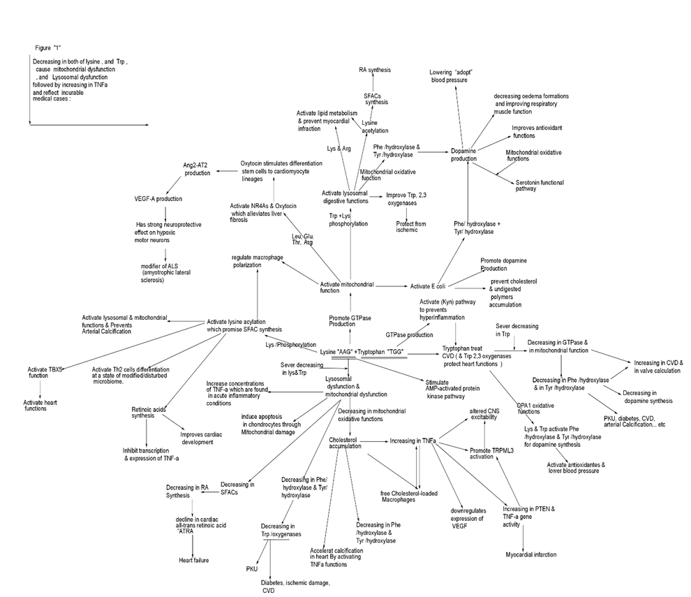
Fig1:
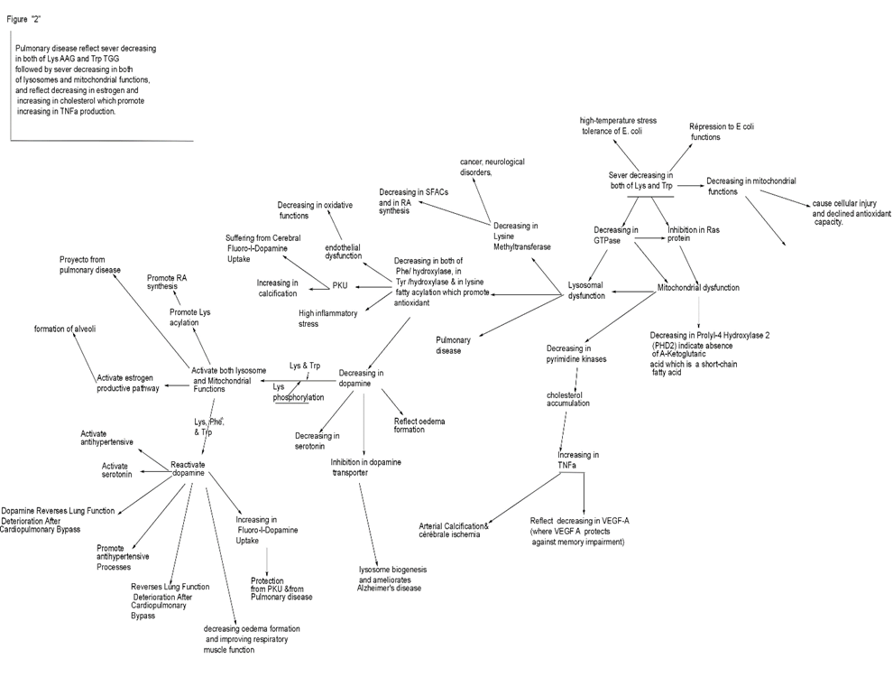
Fig2
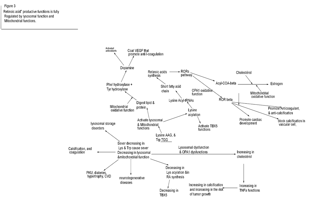
Fig3:
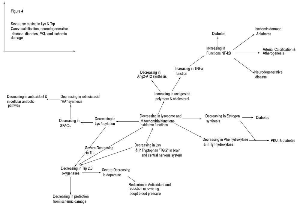
Fig4:
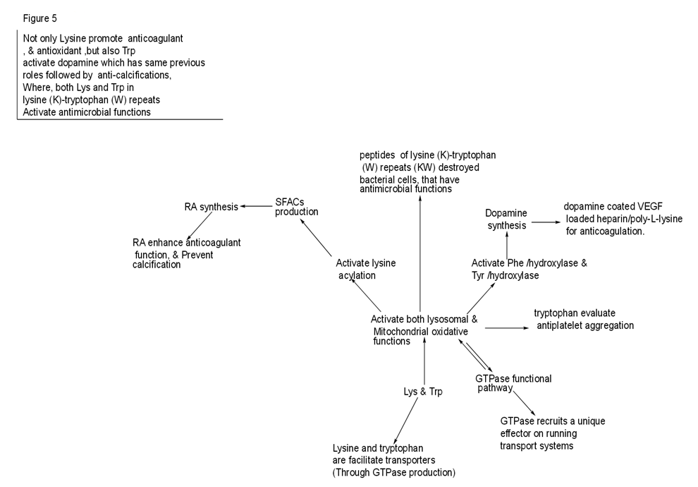
Fig5:
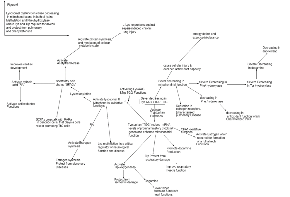
Fig6:
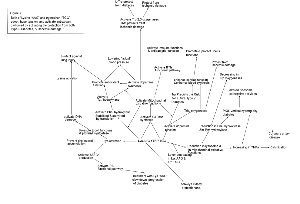
Fig7:
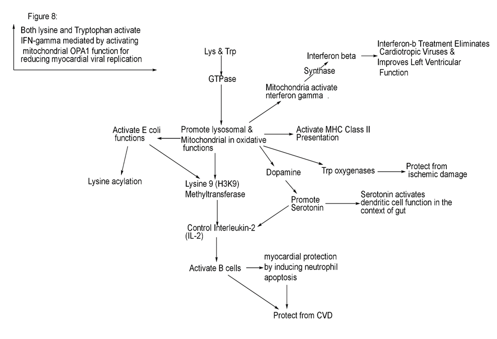
Fig8:
Conclusion
Phenylketonuria characterized by low utilization of GTPase and high accumulated undigested polymers and cholesterol (which activate TNFa pathogenic function), with low antioxidants, mediated by decreased in lysine and tryptophan, and decreasing in both of Phe /hydroxylase and Tyr/ hydroxylase and followed by decreasing in dopamine. Reduction in Trp “TGG” and in Lys “AAG” cause decreasing in oxygen species and in estrogen Synthesis (mediated by decreasing in Mitochondrial region and function), followed by accumulation in androgen and in pro-inflammation including cholesterol which activate the increasing in TNFa functional pathways.
-Lysosomes as important native tools for controlling B cells function through activating dendritic cells for activating Anti-Tumors, and for activating myocardial protection, that Lys phosphorylation and Trp are having strong roles in activating all of lysosomal function, Mitochondrial oxidative function and E coli functions.
Severe Decreasing in both Lys and Trp will cause sever decreasing in lysosome and Mitochondrial functions followed by cholesterol accumulation and decreasing in Trp oxygenases production, which followed by increasing in TNFa that will cause diabetes, coronary artery disease, ischemic damage, and atherogenesis.
Both Lys and Trp facilitate the amino acid transporters across cells membranes that activate antibacterial function and anti-atherosclerosis, mediated by GTPase productive functions.
the lysosomal dysfunction is Abundant Source of the accumulation of undigested polymers and cholesterol which are the Abundant Source of TNFa, and consequently are the Abundant Source of not normal macrophages and then are the basic source of calcification, CVD, and heart failure.
treatment with lysine and Trp in proper arranged molecular structure will reduce diabetes and phenylketonuria syndrome mediated by activating both of lysosomes and mitochondrial functions followed by activating E coli function and followed by activating nrf2, Angiotensin2 and dopamine productive pathways.
The lysosomal dysfunction promise accumulated phenylalanine, inflammation, and cholesterol deposit in arteries walls and exert calcification because of the accumulation of undigested polymers which included cholesterol and pro-inflammation, followed by increasing in TNFa production al pathway.
The lysosomal dysfunction cause accumulation in the undigested inflammation, polymers, and in the accumulation of cellular waste, that also reflect the cholesterol accumulation.
Lysosome dysfunction contributes to cardiovascular disorders, neurodegenerativee diseases, coronary calcification, and atherogenesis.
Lysine “AAG” has strong role in activating ATPase production which activate lysine phosphorylation in lysosomes, and activate lysine acylation which activate short fatty acid chains which are very necessary for activating retinoic acid functional pathway in vivo which activate antioxidants functions and necessary for activation brain and heart function. Lys and Trp activate Phe hydroxylase which activate E coli functions, followed by activating Tyr/ hydroxylase and dopamine production.
Lysine AAA, AAG, <¬necessary to encode and stabilize the Phe TTT, TTC functions,
and the lysine AAG is the reversed copy of glutamate Glu “GAA”, that are having same functions for activating lysosomes and activating antihypertensive pathway (as discussed before) and both promise the stability of leucine “CTT” functional pathway.
Lysine and Trp activate GTPase which control lysosome function and control both of E coli functions in heart and control Dendritic cell function, and control mitochondrial functions which regulate MHC Class II Presentation (which produced by B cells), that contribute to myocardial protection via activating antihypertensive pathway and NR4As pathway.
Lysine has important role for activating Glu, and leucine and activate GTPase production which necessary for mitochondrial Biogenesis mediated by lysine acetylation production which necessary for activating heart functions.
The absence of lysine AAG and in Trp TGG will cause dysfunction in both of lysosomes and Mitochondrial oxidative functions , followed by decreasing in Phe/ hydroxylase production, and decreasing in lysine acylation (which can be decreasing in RA and antioxidant), that followed by increasing in the accumulation of phenylalanine, and accumulation in both of un-digestive polymers and cholesterol that will cause phenylketonuria associated with diabetes, and also the PKU is characterized by defect in dopamine synthesis and increasing in calcification , with increasing in the risk of blood disorders , cardiac dysfunction, and increase the risk of pulmonary diseases.
Also , we can conclude that both of Lys “AAG” and Trp “TGG ” are so necessary for activating mitochondrial oxidative functions which is so necessary for activating antioxidant functions, which promote Phe/ hydroxylase & Tyr/ hydroxylase production and activate lysine acylation which is so important for activating retinoic acid , where the PKU characterized by sever decreasing in antioxidant function and sever decreasing in Phe/ hydroxylase production.
Also Lys acylation has the importance role in activating short fatty acid chains “SFACs“ and acetyltransferase production which mediated the importance of proper metabolic pathways, and the SFACs has important roles for activating retinoic acid functional pathway, that’s why PKU characterized by reduction in Phe/ hydroxylase, (and reduction in Tyr/ hydroxylase due to reduction in mitochondrial function ) reduction in acetyl transfers, and then reduction in SFACs production followed by reduction in both of retinoic acid function and reduction in dopamine productive functions.
Both lysine and Tryptophan are so necessary for activating mitochondrial repairs and functions for activating IFNs functional pathways, but not the L-12/IL-18.
That Upon availability of both of Lys and Trp functions the Interferon-β will be activated to eliminate Cardiotropic Viruses and Improves Left Ventricular Function.
And, upon availability of Lys and Trp functional pathway the IFN-α and IFN β will be activated for reducing myocardial viral replication and damage.
Conflict of interest
I’m Ashraf M El Tantawi is the only one author that studied and wrote this work. And I am the only author declare that the research was conducted in the absence of any commercial or financial relationships “which could be construed as a potential conflict of interest”.
Author acknowledgment
Thanks, and appreciation to all doctors and professors who presented their work in medical research at highest level, and some of their work has been mentioned in the references of this work.
And, Special thanks to the medical journals who contributed with great effort and work in spreading biomedical sciences, shortening the distances between all doctors and professors, and facilitate distribution the Biomedical articles across universe.
References
- Simonaro, C. M. (2019). Lysosomes, lysosomal storage diseases, and inflammation. Journal of Inborn Errors of Metabolism and Screening, 4, e160023.
- Hou, Y., He, H., Ma, M., & Zhou, R. (2023). Apilimod activates the NLRP3 inflammasome through lysosome-mediated mitochondrial damage. Frontiers in Immunology, 14, 1128700.
- Li, M. M., Wang, X., Chen, X. D., Yang, H. L., Xu, H. S., Zhou, P., ... & Liu, N. (2022). Lysosomal dysfunction is associated with NLRP3 inflammasome activation in chronic unpredictable mild stress-induced depressive mice. Behavioural Brain Research, 432, 113987.
- Murr, C., Grammer, T. B., Meinitzer, A., Kleber, M. E., März, W., & Fuchs, D. (2014). Immune activation and inflammation in patients with cardiovascular disease are associated with higher phenylalanine to tyrosine ratios: the ludwigshafen risk and cardiovascular health study. Journal of amino acids, 2014(1), 783730.
- Cuozzo, J. W., Tao, K., Wu, Q. L., Young, W., & Sahagian, G. G. (1995). Lysine-based Structure in the Proregion of Procathepsin L Is the Recognition Site for Mannose Phosphorylation (∗). Journal of Biological Chemistry, 270(26), 15611-15619.
- Jiang, H., Zhang, X., & Lin, H. (2016). Lysine fatty acylation promotes lysosomal targeting of TNF-α. Scientific reports, 6(1), 24371.
- Shimomura, A., Matsui, I., Hamano, T., Ishimoto, T., Katou, Y., Takehana, K., ... & Rakugi, H. (2014). Dietary L-lysine prevents arterial calcification in adenine-induced uremic rats. Journal of the American Society of Nephrology, 25(9), 1954-1965.
- Riazi, K., Galic, M. A., Kuzmiski, J. B., Ho, W., Sharkey, K. A., & Pittman, Q. J. (2008). Microglial activation and TNFα production mediate altered CNS excitability following peripheral inflammation. Proceedings of the National Academy of Sciences, 105(44), 17151-17156.
- Sato, T., Ito, Y., & Nagasawa, T. (2014). Lysine suppresses myofibrillar protein degradation by regulating the autophagic-lysosomal system through phosphorylation of Akt in C2C12 cells. SpringerPlus, 3, 1-12.
- Tang, K., Ma, J., & Huang, B. (2022). Macrophages’ M1 or M2 by tumor microparticles: lysosome makes decision. Cellular & Molecular Immunology, 19(10), 1196-1197.
- Shimomura, A., Matsui, I., Hamano, T., Ishimoto, T., Katou, Y., Takehana, K., ... & Rakugi, H. (2014). Dietary L-lysine prevents arterial calcification in adenine-induced uremic rats. Journal of the American Society of Nephrology, 25(9), 1954-1965.
- Wang, L., & He, C. (2022). Nrf2-mediated anti-inflammatory polarization of macrophages as therapeutic targets for osteoarthritis. Frontiers in immunology, 13, 967193.
- Miao, Y., Li, G., Zhang, X., Xu, H., & Abraham, S. N. (2015). A TRP channel senses lysosome neutralization by pathogens to trigger their expulsion. Cell, 161(6), 1306-1319.
- Sorgdrager, F. J., Naudé, P. J., Kema, I. P., Nollen, E. A., & Deyn, P. P. D. (2019). Tryptophan metabolism in inflammaging: from biomarker to therapeutic target. Frontiers in immunology, 10, 2565.
- Zhai, X., Zhang, H., Xia, Z., Liu, M., Du, G., Jiang, Z., ... & Jin, B. (2024). Oxytocin alleviates liver fibrosis via hepatic macrophages. JHEP Reports, 6(6), 101032.
- Buemann, B., & Uvnäs-Moberg, K. (2020). Oxytocin may have a therapeutical potential against cardiovascular disease. Possible pharmaceutical and behavioral approaches. Medical hypotheses, 138, 109597.
- Jankowski, M., Broderick, T. L., & Gutkowska, J. (2020). The role of oxytocin in cardiovascular protection. Frontiers in psychology, 11, 2139.
- Mangge, H., Stelzer, I., Reininghaus, E. Z., Weghuber, D., Postolache, T. T., & Fuchs, D. (2014). Disturbed tryptophan metabolism in cardiovascular disease. Current medicinal chemistry, 21(17), 1931-1937.
- Ansari, M. Y., Ball, H. C., Wase, S. J., Novak, K., & Haqqi, T. M. (2021). Lysosomal dysfunction in osteoarthritis and aged cartilage triggers apoptosis in chondrocytes through BAX mediated release of Cytochrome c. Osteoarthritis and cartilage, 29(1), 100-112.
- Astorga, J., Gasaly, N., Dubois-Camacho, K., De la Fuente, M., Landskron, G., Faber, K. N., ... & Hermoso, M. A. (2022). The role of cholesterol and mitochondrial bioenergetics in activation of the inflammasome in IBD. Frontiers in immunology, 13, 1028953.
- Li, Y., Schwabe, R. F., DeVries-Seimon, T., Yao, P. M., Gerbod-Giannone, M. C., Tall, A. R., ... & Tabas, I. (2005). Free cholesterol-loaded macrophages are an abundant source of tumor necrosis factor-α and interleukin-6: model of NF-κB-and map kinase-dependent inflammation in advanced atherosclerosis. Journal of Biological Chemistry, 280(23), 21763-21772.
- Tian, M., Yuan, Y. C., Li, J. Y., Gionfriddo, M. R., & Huang, R. C. (2015). Tumor necrosis factor-α and its role as a mediator in myocardial infarction: A brief review. Chronic diseases and translational medicine, 1(1), 18-26.
- Li, M. M., Wang, X., Chen, X. D., Yang, H. L., Xu, H. S., Zhou, P., ... & Liu, N. (2022). Lysosomal dysfunction is associated with NLRP3 inflammasome activation in chronic unpredictable mild stress-induced depressive mice. Behavioural Brain Research, 432, 113987.
- McGeough, M. D., Wree, A., Inzaugarat, M. E., Haimovich, A., Johnson, C. D., Peña, C. A., ... & Hoffman, H. M. (2017). TNF regulates transcription of NLRP3 inflammasome components and inflammatory molecules in cryopyrinopathies. The Journal of clinical investigation, 127(12), 4488-4497.
- Rolski, F., & Błyszczuk, P. (2020). Complexity of TNF-α signaling in heart disease. Journal of clinical medicine, 9(10), 3267.
- Mahmoud, A. H., Taha, N. M., Zakhary, M., & Tadros, M. S. (2019). PTEN gene & TNF-alpha in acute myocardial infarction. IJC Heart & Vasculature, 23, 100366.
- Ebenezar, K. K., Sathish, V., & Devaki, T. (2003). Effect of oral administration of L-arginine and L-lysine on lipid metabolism against isoproterenol-induced myocardial infarction in rats. Journal of Clinical Biochemistry and Nutrition, 33(1), 7-11.
- Wal, P., Aziz, N., Singh, Y. K., Wal, A., Kosey, S., & Rai, A. K. (2023). Myocardial infarction as a consequence of mitochondrial dysfunction. Current Cardiology Reviews, 19(6), 23-30.
- Curtius, H. C., Niederwieser, A., Viscontini, M., Leimbacher, W., Wegmann, H., Blehova, B., ... & Schmidt, H. (1981). Serotonin and dopamine synthesis in phenylketonuria. Serotonin: Current aspects of neurochemistry and function, 277-291.
- Rodriguez, A. G., Schroeder, M. E., Grim, J. C., Walker, C. J., Speckl, K. F., Weiss, R. M., & Anseth, K. S. (2021). Tumor necrosis factor-α promotes and exacerbates calcification in heart valve myofibroblast populations. FASEB journal: official publication of the Federation of American Societies for Experimental Biology, 35(3), e21382.
- Padfield, G. J., Din, J. N., Koushiappi, E., Mills, N. L., Robinson, S. D., Cruden, N. L. M., ... & Newby, D. E. (2013). Cardiovascular effects of tumour necrosis factor α antagonism in patients with acute myocardial infarction: a first in human study. Heart, 99(18), 1330-1335.
- Popa, C., Netea, M. G., Van Riel, P. L., Van Der Meer, J. W., & Stalenhoef, A. F. (2007). The role of TNF-α in chronic inflammatory conditions, intermediary metabolism, and cardiovascular risk. Journal of lipid research, 48(4), 751-762.
- Yuan, S., Carter, P., Bruzelius, M., Vithayathil, M., Kar, S., Mason, A. M., ... & Larsson, S. C. (2020). Effects of tumour necrosis factor on cardiovascular disease and cancer: A two-sample Mendelian randomization study. EBioMedicine, 59.
- Shimomura, A., Matsui, I., Hamano, T., Ishimoto, T., Katou, Y., Takehana, K., ... & Rakugi, H. (2014). Dietary L-lysine prevents arterial calcification in adenine-induced uremic rats. Journal of the American Society of Nephrology, 25(9), 1954-1965.
- Zhao, R., Cai, K., Yang, J. J., Zhou, Q., Cao, W., Xiang, J., ... & Zhao, J. Y. (2023). Nuclear ATR lysine-tyrosylation protects against heart failure by activating DNA damage response. Cell Reports, 42(4).
- Chen, S. Y., Lin, W. C., Chang, Y. Y., Lin, T. K., & Lan, M. Y. (2020). Brain hypoperfusion and nigrostriatal dopaminergic dysfunction in primary familial brain calcification caused by novel MYORG variants: case report. BMC neurology, 20, 1-6.
- Shimomura, A., Matsui, I., Hamano, T., Ishimoto, T., Katou, Y., Takehana, K., ... & Rakugi, H. (2014). Dietary L-lysine prevents arterial calcification in adenine-induced uremic rats. Journal of the American Society of Nephrology, 25(9), 1954-1965.
- Reichard, A., & Asosingh, K. (2019). The role of mitochondria in angiogenesis. Molecular biology reports, 46, 1393-1400.
- Takahashi, H., & Shibuya, M. (2005). The vascular endothelial growth factor (VEGF)/VEGF receptor system and its role under physiological and pathological conditions. Clinical science, 109(3), 227-241.
- Qi, F., Zuo, Z., Hu, K., Wang, R., Wu, T., Liu, H., ... & Guo, K. (2023). VEGF-A in serum protects against memory impairment in APP/PS1 transgenic mice by blocking neutrophil infiltration. Molecular Psychiatry, 28(10), 4374-4389.
- Tantawi, A. E. (2023). DPP4 valorphin activate NR4As pathway and OPA1 that protect from CoQ10 deficien-cy from OPA1 dysfunctions and from WMH. J Pharmaceut Res, 8(2), 271-300.
- Chen, X., Austin, E. D., Talati, M., Fessel, J. P., Farber-Eger, E. H., Brittain, E. L., ... & West, J. (2017). Oestrogen inhibition reverses pulmonary arterial hypertension and associated metabolic defects. European Respiratory Journal, 50(2).
- Pokharel, M. D., Garcia-Flores, A., Marciano, D., Franco, M. C., Fineman, J. R., Aggarwal, S., ... & Black, S. M. (2024). Mitochondrial network dynamics in pulmonary disease: Bridging the gap between inflammation, oxidative stress, and bioenergetics. Redox Biology, 103049.
- Ryter, S. W., Rosas, I. O., Owen, C. A., Martinez, F. J., Choi, M. E., Lee, C. G., ... & Choi, A. M. (2018). Mitochondrial dysfunction as a pathogenic mediator of chronic obstructive pulmonary disease and idiopathic pulmonary fibrosis. Annals of the American Thoracic Society, 15(Supplement 4), S266-S272.
- Dai, Z., Li, M., Wharton, J., Zhu, M. M., & Zhao, Y. Y. (2016). Prolyl-4 hydroxylase 2 (PHD2) deficiency in endothelial cells and hematopoietic cells induces obliterative vascular remodeling and severe pulmonary arterial hypertension in mice and humans through hypoxia-inducible factor-2α. Circulation, 133(24), 2447-2458.
- Wong, Y. C., Kim, S., Peng, W., & Krainc, D. (2019). Regulation and function of mitochondria–lysosome membrane contact sites in cellular homeostasis. Trends in cell biology, 29(6), 500-513.
- Hlavata, L., & Nyström, T. (2003). Ras proteins control mitochondrial biogenesis and function in Saccharomyces cerevisiae. Folia microbiologica, 48, 725-730.
- Fernandes, J., Weddle, A., Kinter, C. S., Humphries, K. M., Mather, T., Szweda, L. I., & Kinter, M. (2015). Lysine acetylation activates mitochondrial aconitase in the heart. Biochemistry, 54(25), 4008-4018.
- Feoli, A., Viviano, M., Cipriano, A., Milite, C., Castellano, S., & Sbardella, G. (2022). Lysine methyltransferase inhibitors: where we are now. RSC Chemical Biology, 3(4), 359-406.
- Bagchi, R. A., Robinson, E. L., Hu, T., Cao, J., Hong, J. Y., Tharp, C. A., ... & McKinsey, T. A. (2022). Reversible lysine fatty acylation of an anchoring protein mediates adipocyte adrenergic signaling. Proceedings of the National Academy of Sciences, 119(7), e2119678119.
- Ghosh, T. K., Aparicio-Sánchez, J. J., Buxton, S., Ketley, A., Mohamed, T., Rutland, C. S., ... & Brook, J. D. (2018). Acetylation of TBX5 by KAT2B and KAT2A regulates heart and limb development. Journal of Molecular and Cellular Cardiology, 114, 185-198.
- Jiao, J., Xia, Y., Zhang, Y., Wu, X., Liu, C., Feng, J., ... & Pang, H. (2021). Phenylalanine 4-hydroxylase contributes to endophytic bacterium Pseudomonas fluorescens’ melatonin biosynthesis. Frontiers in Genetics, 12, 746392.
- Rattazzi, M., Donato, M., Bertacco, E., Millioni, R., Franchin, C., Mortarino, C., ... & Arrigoni, G. (2020). l-Arginine prevents inflammatory and pro-calcific differentiation of interstitial aortic valve cells. Atherosclerosis, 298, 27-35.
- Rodionov, R. N., Begmatov, H., Jarzebska, N., Patel, K., Mills, M. T., Ghani, Z., ... & Savinova, O. V. (2019). Homoarginine supplementation prevents left ventricular dilatation and preserves systolic function in a model of coronary artery disease. Journal of the American Heart Association, 8(14), e012486.
- Azabdaftari, A., van der Giet, M., Schuchardt, M., Hennermann, J. B., Plöckinger, U., & Querfeld, U. (2019). The cardiovascular phenotype of adult patients with phenylketonuria. Orphanet journal of rare diseases, 14, 1-11.
- Woodring, J. H., & Rosenbaum, H. D. (1981). Bone changes in phenylketonuria reassessed. American Journal of Roentgenology, 137(2), 241-243.
- Gudinchet, F., Maeder, P. H., Meuli, R. A., Deonna, T. H., & Mathieu, J. M. (1992). Cranial CT and MRI in malignant phenylketonuria. Pediatric radiology, 22, 223-224.
- El Tantawi, A. M. (2022). DCs-IL2 Necessary for Glucocorticoids Which Necessary for Interferons Synthesis and Serotonin Synthesis then Promote Ang2-At2 and VEGF-A for Anti-Inflammatory Growth. Stem Cells Regen Med, 6(1), 1-27.
- Bai, Y., Wang, H. M., Liu, M., Wang, Y., Lian, G. C., Zhang, X. H., ... & Wang, H. L. (2014). 4-Chloro-DL-phenylalanine protects against monocrotaline induced pulmonary vascular remodeling and lung inflammation. International journal of molecular medicine, 33(2), 373-382.
- Liu, B., Du, H., Rutkowski, R., Gartner, A., & Wang, X. (2012). LAAT-1 is the lysosomal lysine/arginine transporter that maintains amino acid homeostasis. Science, 337(6092), 351-354.
- Cuozzo, J. W., Tao, K., Wu, Q. L., Young, W., & Sahagian, G. G. (1995). Lysine-based Structure in the Proregion of Procathepsin L Is the Recognition Site for Mannose Phosphorylation (∗). Journal of Biological Chemistry, 270(26), 15611-15619.
- Sirtori, L. R., Dutra-Filho, C. S., Fitarelli, D., Sitta, A., Haeser, A., Barschak, A. G., ... & Vargas, C. R. (2005). Oxidative stress in patients with phenylketonuria. Biochimica et Biophysica Acta (BBA)-Molecular Basis of Disease, 1740(1), 68-73.
- Dobrowolski, S. F., Phua, Y. L., Vockley, J., Goetzman, E., & Blair, H. C. (2022). Phenylketonuria oxidative stress and energy dysregulation: emerging pathophysiological elements provide interventional opportunity. Molecular genetics and metabolism, 136(2), 111-117.
- Bhatti, J. S., Bhatti, G. K., & Reddy, P. H. (2017). Mitochondrial dysfunction and oxidative stress in metabolic disorders—A step towards mitochondria based therapeutic strategies. Biochimica et Biophysica Acta (BBA)-Molecular Basis of Disease, 1863(5), 1066-1077.
- Jiang, Q., Yin, J., Chen, J., Ma, X., Wu, M., Liu, G., ... & Yin, Y. (2020). Mitochondria‐targeted antioxidants: a step towards disease treatment. Oxidative medicine and cellular longevity, 2020(1), 8837893.
- Jiang, Q., Yin, J., Chen, J., Ma, X., Wu, M., Liu, G., ... & Yin, Y. (2020). Mitochondria‐targeted antioxidants: a step towards disease treatment. Oxidative medicine and cellular longevity, 2020(1), 8837893.
- Liu, G., Sun, W., Wang, F., Jia, G., Zhao, H., Chen, X., ... & Wang, J. (2023). Dietary tryptophan supplementation enhances mitochondrial function and reduces pyroptosis in the spleen and thymus of piglets after lipopolysaccharide challenge. animal, 17(3), 100714.
- Xie, S., He, J., Masagounder, K., Liu, Y., Tian, L., Tan, B., & Niu, J. (2022). Dietary lysine levels modulate the lipid metabolism, mitochondrial biogenesis and immune response of grass carp, Ctenopharyngodon idellus. Animal Feed Science and Technology, 291, 115375.
- Fernandes, J., Weddle, A., Kinter, C. S., Humphries, K. M., Mather, T., Szweda, L. I., & Kinter, M. (2015). Lysine acetylation activates mitochondrial aconitase in the heart. Biochemistry, 54(25), 4008-4018.
- Thomas, S. P., & Denu, J. M. (2021). Short-chain fatty acids activate acetyltransferase p300. Elife, 10, e72171.
- Blasl, A. T., Schulze, S., Qin, C., Graf, L. G., Vogt, R., & Lammers, M. (2022). Post-translational lysine ac (et) ylation in health, ageing and disease. Biological Chemistry, 403(2), 151-194.
- Yuan, X., Tang, H., Wu, R., Li, X., Jiang, H., Liu, Z., & Zhang, Z. (2021). Short-chain fatty acids calibrate RARα activity regulating food sensitization. Frontiers in Immunology, 12, 737658.
- Black, J. C., Van Rechem, C., & Whetstine, J. R. (2012). Histone lysine methylation dynamics: establishment, regulation, and biological impact. Molecular cell, 48(4), 491-507.
- Ciarka, A., Vincent, J. L., & Van de Borne, P. (2007). The effects of dopamine on the respiratory system: friend or foe?. Pulmonary pharmacology & therapeutics, 20(6), 607-615.
- Peták, F., Balogh, Á. L., Hankovszky, P., Fodor, G. H., Tolnai, J., Südy, R., ... & Babik, B. (2022). Dopamine Reverses Lung Function Deterioration After Cardiopulmonary Bypass Without Affecting Gas Exchange. Journal of Cardiothoracic and Vascular Anesthesia, 36(4), 1047-1055.
- Massaro, D., & Massaro, G. D. (2004). Estrogen regulates pulmonary alveolar formation, loss, and regeneration in mice. American journal of physiology-lung cellular and molecular physiology, 287(6), L1154-L1159.
- Frost, F., Fothergill, J., Winstanley, C., Nazareth, D., & Walshaw, M. J. (2019). S20 Inhaled aztreonam lysine recovers lung function and improves quality of life in acute pulmonary exacerbations of cystic fibrosis.
- McAdams, R. M., & Traudt, C. M. (2018). Brain injury in the term infant. In Avery's Diseases of the Newborn (pp. 897-909). Elsevier.
- Landvogt, C., Mengel, E., Bartenstein, P., Buchholz, H. G., Schreckenberger, M., Siessmeier, T., ... & Ullrich, K. (2008). Reduced cerebral fluoro-L-dopamine uptake in adult patients suffering from phenylketonuria. Journal of Cerebral Blood Flow & Metabolism, 28(4), 824-831.
- Vockley, J., Andersson, H. C., Antshel, K. M., Braverman, N. E., Burton, B. K., Frazier, D. M., ... & Berry, S. A. (2014). Phenylalanine hydroxylase deficiency: diagnosis and management guideline. Genetics in medicine, 16(2), 188-200.
- Mishra, A., Singh, S., Tiwari, V., Chaturvedi, S., Wahajuddin, M., & Shukla, S. (2019). Dopamine receptor activation mitigates mitochondrial dysfunction and oxidative stress to enhance dopaminergic neurogenesis in 6-OHDA lesioned rats: A role of Wnt signalling. Neurochemistry international, 129, 104463.
- Neumann‐Staubitz, P., Lammers, M., & Neumann, H. (2021). Genetic code expansion tools to study lysine acylation. Advanced Biology, 5(12), 2100926.
- Yin, L., Zhou, J., Li, T., Wang, X., Xue, W., Zhang, J., ... & Li, Y. (2023). Inhibition of the dopamine transporter promotes lysosome biogenesis and ameliorates Alzheimer's disease–like symptoms in mice. Alzheimer's & Dementia, 19(4), 1343-1357.
- Chen, W. S., Wang, C. H., Cheng, C. W., Liu, M. H., Chu, C. M., Wu, H. P., ... & Liang, C. Y. (2020). Elevated plasma phenylalanine predicts mortality in critical patients with heart failure. ESC Heart failure, 7(5), 2884-2893.
- Zhao, R., Cai, K., Yang, J. J., Zhou, Q., Cao, W., Xiang, J., ... & Zhao, J. Y. (2023). Nuclear ATR lysine-tyrosylation protects against heart failure by activating DNA damage response. Cell Reports, 42(4).
- Isobe, K., Matsui, D., & Asano, Y. (2019). Comparative review of the recent enzymatic methods used for selective assay of L-lysine. Analytical biochemistry, 584, 113335.
- Daubner, S. C., Le, T., & Wang, S. (2011). Tyrosine hydroxylase and regulation of dopamine synthesis. Archives of biochemistry and biophysics, 508(1), 1-12.
- Lou, H. C. (1994). Dopamine precursors and brain function in phenylalanine hydroxylase deficiency. Acta Paediatrica, 83, 86-88.
- Bueno-Carrasco, M. T., Cuéllar, J., Flydal, M. I., Santiago, C., Kråkenes, T. A., Kleppe, R., ... & Valpuesta, J. M. (2022). Structural mechanism for tyrosine hydroxylase inhibition by dopamine and reactivation by Ser40 phosphorylation. Nature communications, 13(1), 74.
- Daubner, S. C., Le, T., & Wang, S. (2011). Tyrosine hydroxylase and regulation of dopamine synthesis. Archives of biochemistry and biophysics, 508(1), 1-12.
- Tan, R., Li, J., Liu, F., Liao, P., Ruiz, M., Dupuis, J., ... & Hu, Q. (2020). Phenylalanine induces pulmonary hypertension through calcium-sensing receptor activation. American Journal of Physiology-Lung Cellular and Molecular Physiology, 319(6), L1010-L1020.
- Neumann, J., Hofmann, B., Dhein, S., & Gergs, U. (2023). Role of Dopamine in the Heart in Health and Disease. International Journal of Molecular Sciences, 24(5), 5042.
- Goldberg, L. I. (1984). Dopamine receptors and hypertension: Physiologic and pharmacologic implications. The American journal of medicine, 77(4), 37-44.
- Zeng, C., & Jose, P. A. (2011). Dopamine receptors: important antihypertensive counterbalance against hypertensive factors. Hypertension, 57(1), 11-17.
- Drake, A. J., Templeton, W. W., & Stanford, S. C. (1982). Evidence for a role of dopamine in cardiac function. Neurochemistry International, 4(5), 435-439.
- Severyanova, L. A., Lazarenko, V. A., Plotnikov, D. V., Dolgintsev, M. E., & Kriukov, A. A. (2019). L-lysine as the molecule influencing selective brain activity in pain-induced behavior of rats. International Journal of Molecular Sciences, 20(8), 1899.
- Bucolo, C., Leggio, G. M., Drago, F., & Salomone, S. (2019). Dopamine outside the brain: The eye, cardiovascular system and endocrine pancreas. Pharmacology & Therapeutics, 203, 107392.
- Azabdaftari, A., van der Giet, M., Schuchardt, M., Hennermann, J. B., Plöckinger, U., & Querfeld, U. (2019). The cardiovascular phenotype of adult patients with phenylketonuria. Orphanet journal of rare diseases, 14, 1-11.
- Jiang, Y., Zhou, J., Zhao, J., Hou, D., Zhang, H., Li, L., ... & Jing, Z. (2020). MiR-18a-downregulated RORA inhibits the proliferation and tumorigenesis of glioma using the TNF-α-mediated NF-κB signaling pathway. EBioMedicine, 52.
- Qu, S., Gao, Y., Ma, J., & Yan, Q. (2023). Microbiota-derived short-chain fatty acids functions in the biology of B lymphocytes: From differentiation to antibody formation. Biomedicine & Pharmacotherapy, 168, 115773.
- Lin, S. C., Dollé, P., Ryckebüsch, L., Noseda, M., Zaffran, S., Schneider, M. D., & Niederreither, K. (2010). Endogenous retinoic acid regulates cardiac progenitor differentiation. Proceedings of the National Academy of Sciences, 107(20), 9234-9239.
- St. Hilaire, C. (2020). Retinoids: dissolving the calcification paradox. Arteriosclerosis, thrombosis, and vascular biology, 40(3), 503-505.
- Yang, N., Parker, L. E., Yu, J., Jones, J. W., Liu, T., Papanicolaou, K. N., ... & Foster, D. B. (2021). Cardiac retinoic acid levels decline in heart failure. JCI insight, 6(8).
- Wu, Y., Huang, T., Li, X., Shen, C., Ren, H., Wang, H., ... & Cai, W. (2023). Retinol dehydrogenase 10 reduction mediated retinol metabolism disorder promotes diabetic cardiomyopathy in male mice. Nature Communications, 14(1), 1181.
- Rogers, M. A., Chen, J., Nallamshetty, S., Pham, T., Goto, S., Muehlschlegel, J. D., ... & Plutzky, J. (2020). Retinoids repress human cardiovascular cell calcification with evidence for distinct selective retinoid modulator effects. Arteriosclerosis, thrombosis, and vascular biology, 40(3), 656-669.
- Miao, Y., Li, G., Zhang, X., Xu, H., & Abraham, S. N. (2015). A TRP channel senses lysosome neutralization by pathogens to trigger their expulsion. Cell, 161(6), 1306-1319.
- Sato, A., Endo, Y., & Natori, Y. (1992). Involvement of lysosomes in substrate stabilization of tryptophan-2, 3-dioxygenase in rat liver. Biochemical and biophysical research communications, 183(1), 306-311.
- Bigelman, E., Pasmanik-Chor, M., Dassa, B., Itkin, M., Malitsky, S., Dorot, O., ... & Entin-Meer, M. (2023). Kynurenic acid, a key L-tryptophan-derived metabolite, protects the heart from an ischemic damage. PLoS One, 18(8), e0275550.
- Bandyopadhyay, U., Todorova, P., Pavlova, N. N., Tada, Y., Thompson, C. B., Finley, L. W., & Overholtzer, M. (2022). Leucine retention in lysosomes is regulated by starvation. Proceedings of the National Academy of Sciences, 119(6), e2114912119.
- Raehtz, S., Bierhalter, H., Schoenherr, D., Parameswaran, N., & McCabe, L. R. (2017). Estrogen deficiency exacerbates type 1 diabetes–induced bone tnf-α expression and osteoporosis in female mice. Endocrinology, 158(7), 2086-2101.
- Couto, R. D., Dallan, L. A., Lisboa, L. A., Mesquita, C. H., Vinagre, C. G., & Maranhão, R. C. (2007). Deposition of free cholesterol in the blood vessels of patients with coronary artery disease: a possible novel mechanism for atherogenesis. Lipids, 42, 411-418.
- Marques, A. R., Ramos, C., Machado-Oliveira, G., & Vieira, O. V. (2021). Lysosome (Dys) function in atherosclerosis—a big weight on the shoulders of a small organelle. Frontiers in cell and developmental biology, 9, 658995.
- Chi, C., Zhu, H., Han, M., Zhuang, Y., Wu, X., & Xu, T. (2010). Disruption of lysosome function promotes tumor growth and metastasis in Drosophila. Journal of Biological Chemistry, 285(28), 21817-21823.
- Root, J., Merino, P., Nuckols, A., Johnson, M., & Kukar, T. (2021). Lysosome dysfunction as a cause of neurodegenerative diseases: Lessons from frontotemporal dementia and amyotrophic lateral sclerosis. Neurobiology of disease, 154, 105360.
- Chi, C., Riching, A. S., & Song, K. (2020). Lysosomal abnormalities in cardiovascular disease. International journal of molecular sciences, 21(3), 811.
- Liu, Y., Zhang, J., Wang, J., Wang, Y., Zeng, Z., Liu, T., ... & Huang, N. (2015). Tailoring of the dopamine coated surface with VEGF loaded heparin/poly‐l‐lysine particles for anticoagulation and accelerate in situ endothelialization. Journal of Biomedical Materials Research Part A, 103(6), 2024-2034.
- Shimomura, A., Matsui, I., Hamano, T., Ishimoto, T., Katou, Y., Takehana, K., ... & Rakugi, H. (2014). Dietary L-lysine prevents arterial calcification in adenine-induced uremic rats. Journal of the American Society of Nephrology, 25(9), 1954-1965.
- Tantawi, A. M. E. (2024). Decreasing in Lysine Reflect Lysosomal Dysfunction and Accumulated Phenylalanine which Connected to WMH and CVD and Both of Phenylke-tonuria and Calcifications where N-Acetylcysteine Prevent Organs Failure. Clin Onco, 7(11), 1-28.
- Xie, Z., Feng, S., Wang, Y., Cao, C., Huang, J., Chen, Y., ... & Li, Z. (2015). Design, synthesis of novel tryptophan derivatives for antiplatelet aggregation activity based on tripeptide pENW (pGlu-Asn-Trp). European journal of medicinal chemistry, 102, 363-374.
- Ogawa, M., Shimizu, F., Ishii, Y., Takao, T., & Takada, A. (2023). Uniqueness of tryptophan in the transport system in the brain and peripheral tissues. Food and Nutrition Sciences, 14(5), 401-414.
- Li, Q., Yang, C., Feng, L., Zhao, Y., Su, Y., Liu, H., ... & Wang, X. (2021). Glutaric acidemia, pathogenesis and nutritional therapy. Frontiers in Nutrition, 8, 704984.
- Liaqat, R., Fatima, S., Komal, W., Minahal, Q., & Hussain, A. S. (2024). Dietary supplementation of methionine, lysine, and tryptophan as possible modulators of growth, immune response, and disease resistance in striped catfish (Pangasius hypophthalmus). Plos one, 19(4), e0301205.
- Gopal, R., Seo, C. H., Song, P. I., & Park, Y. (2013). Effect of repetitive lysine–tryptophan motifs on the bactericidal activity of antimicrobial peptides. Amino acids, 44, 645-660.
- Nitz, K., Lacy, M., & Atzler, D. (2019). Amino acids and their metabolism in atherosclerosis. Arteriosclerosis, thrombosis, and vascular biology, 39(3), 319-330.
- Mizuno-Yamasaki, E., Rivera-Molina, F., & Novick, P. (2012). GTPase networks in membrane traffic. Annual review of biochemistry, 81(1), 637-659.
- Ciarka, A., Vincent, J. L., & Van de Borne, P. (2007). The effects of dopamine on the respiratory system: friend or foe?. Pulmonary pharmacology & therapeutics, 20(6), 607-615.
- Gulcev, M., Reilly, C., Griffin, T. J., Broeckling, C. D., Sandri, B. J., Witthuhn, B. A., ... & Wendt, C. H. (2016). Tryptophan catabolism in acute exacerbations of chronic obstructive pulmonary disease. International journal of chronic obstructive pulmonary disease, 2435-2446.
- Gray, H. B., & Winkler, J. R. (2015). Hole hopping through tyrosine/tryptophan chains protects proteins from oxidative damage. Proceedings of the National Academy of Sciences, 112(35), 10920-10925.
- Li, X. W., Lin, Y. Z., Lin, H., Huang, J. B., Tang, X. M., Long, X. M., ... & Zhao, X. F. (2016). Histidine-tryptophan-ketoglutarate solution decreases mortality and morbidity in high-risk patients with severe pulmonary arterial hypertension associated with complex congenital heart disease: an 11-year experience from a single institution. Brazilian Journal of Medical and Biological Research, 49(6), e5208.
- Kawahara, A., & Stainier, D. Y. (2009). Noncanonical activity of seryl-transfer RNA synthetase and vascular development. Trends in cardiovascular medicine, 19(6), 179-182.
- Li, Z., Siddique, I., Hadrović, I., Kirupakaran, A., Li, J., Zhang, Y., ... & Bitan, G. (2021). Lysine-selective molecular tweezers are cell penetrant and concentrate in lysosomes. Communications biology, 4(1), 1076.
- Zhang, C., He, Y., & Shen, Y. (2019). L-Lysine protects against sepsis-induced chronic lung injury in male albino rats. Biomedicine & Pharmacotherapy, 117, 109043.
- Jiang, H., Zhang, X., & Lin, H. (2016). Lysine fatty acylation promotes lysosomal targeting of TNF-α. Scientific reports, 6(1), 24371.
- Noritsugu, K., Suzuki, T., Dodo, K., Ohgane, K., Ichikawa, Y., Koike, K., ... & Ito, A. (2023). Lysine long-chain fatty acylation regulates the TEAD transcription factor. Cell Reports, 42(4).
- Liu, B., Du, H., Rutkowski, R., Gartner, A., & Wang, X. (2012). LAAT-1 is the lysosomal lysine/arginine transporter that maintains amino acid homeostasis. Science, 337(6092), 351-354.
- Arines, F. M., Wielenga, A., Henn, D., Burata, O. E., Garcia, F. N., Stockbridge, R. B., & Li, M. (2024). Lysosomal membrane transporter purification and reconstitution for functional studies. Molecular Biology of the Cell, 35(3), ar28.
- Dong, X. P., Shen, D., Wang, X., Dawson, T., Li, X., Zhang, Q., ... & Xu, H. (2010). PI (3, 5) P2 controls membrane trafficking by direct activation of mucolipin Ca2+ release channels in the endolysosome. Nature communications, 1(1), 38.
- Borisovska, M., Schwarz, Y. N., Dhara, M., Yarzagaray, A., Hugo, S., Narzi, D., ... & Bruns, D. (2012). Membrane-proximal tryptophans of synaptobrevin II stabilize priming of secretory vesicles. Journal of Neuroscience, 32(45), 15983-15997.
- Rodrı́guez-Alfaro, J. A., Gomez-Fernandez, J. C., & Corbalan-Garcia, S. (2004). Role of the lysine-rich cluster of the C2 domain in the phosphatidylserine-dependent activation of PKCα. Journal of molecular biology, 335(4), 1117-1129.
- Guglielmelli, A., Bartucci, R., Rizzuti, B., Palermo, G., Guzzi, R., & Strangi, G. (2023). The interaction of tryptophan enantiomers with model membranes is modulated by polar head type and physical state of phospholipids. Colloids and Surfaces B: Biointerfaces, 224, 113216.
- Lee, J. Y., Kim, N. A., Sanford, A., & Sullivan, K. E. (2003). Histone acetylation and chromatin conformation are regulated separately at the TNF-α promoter in monocytes and macrophages. Journal of Leucocyte Biology, 73(6), 862-871.
- Rahman, I., Gilmour, P. S., Jimenez, L. A., & MacNee, W. (2002). Oxidative stress and TNF-a induce histone Acetylation and NF-кB/AP-1 activation in Alveolar epithelial cells: Potential mechanism In gene transcription in lung inflammation. Oxygen/Nitrogen Radicals: Cell Injury and Disease, 239-248.
- Davis, F. M., Joshi, A. D., Wolf, S. J., Allen, R., Lipinski, J., Nguyen, B., ... & Gallagher, K. A. (2020). TNF-α regulates diabetic macrophage function through the histone acetyltransferase MOF. JCI insight, 5(5).
- Touyz, R. M., Deng, L. Y., He, G., Wu, X. H., & Schiffrin, E. L. (1999). Angiotensin II stimulates DNA and protein synthesis in vascular smooth muscle cells from human arteries: role of extracellular signal-regulated kinases. Journal of hypertension, 17(7), 907-916.
- Ibrahim, J., Hughes, A. D., & Sever, P. S. (2000). Action of angiotensin II on DNA synthesis by human saphenous vein in organ culture. Hypertension, 36(5), 917-921.
- Guo, L., Tian, H., Shen, J., Zheng, C., Liu, S., Cao, Y., ... & Yao, J. (2018). Phenylalanine regulates initiation of digestive enzyme mRNA translation in pancreatic acinar cells and tissue segments in dairy calves. Bioscience Reports, 38(1), BSR20171189.
- Ramasubbu, N., Ragunath, C., Mishra, P. J., Thomas, L. M., Gyémánt, G., & Kandra, L. (2004). Human salivary α‐amylase Trp58 situated at subsite− 2 is critical for enzyme activity. European Journal of Biochemistry, 271(12), 2517-2529.
- Pati, D., & Kash, T. L. (2021). Tumor necrosis factor-α modulates GABAergic and dopaminergic neurons in the ventrolateral periaqueductal gray of female mice. Journal of Neurophysiology, 126(6), 2119-2129.
- van Heesch, F., Prins, J., Korte-Bouws, G. A., Westphal, K. G., Lemstra, S., Olivier, B., ... & Korte, S. M. (2013). Systemic tumor necrosis factor-alpha decreases brain stimulation reward and increases metabolites of serotonin and dopamine in the nucleus accumbens of mice. Behavioural brain research, 253, 191-195.
- Abe, M., Iwaoka, M., Nakamura, T., Kitta, Y., Takano, H., Kodama, Y., ... & Kugiyama, K. (2007). Association of high levels of plasma free dopamine with future coronary events in patients with coronary artery disease. Circulation Journal, 71(5), 688-692.
- Jiang, H., Zhang, X., & Lin, H. (2016). Lysine fatty acylation promotes lysosomal targeting of TNF-α. Scientific reports, 6(1), 24371.
- Huang, T. L., Wu, C. C., Yu, J., Sumi, S., & Yang, K. C. (2016). l-Lysine regulates tumor necrosis factor-alpha and matrix metalloproteinase-3 expression in human osteoarthritic chondrocytes. Process Biochemistry, 51(7), 904-911.
- Owens, P., & O’Brien, E. (1999). Hypotension in patients with coronary disease: can profound hypotensive events cause myocardial ischaemic events?. Heart, 82(4), 477-481.
- Iellamo, F., Perrone, M. A., Caminiti, G., Volterrani, M., & Legramante, J. M. (2021). Post-exercise Hypotension in Patients With Coronary Artery Disease. Frontiers in Physiology, 12, 788591.
- Shimomura, A., Matsui, I., Hamano, T., Ishimoto, T., Katou, Y., Takehana, K., ... & Rakugi, H. (2014). Dietary L-lysine prevents arterial calcification in adenine-induced uremic rats. Journal of the American Society of Nephrology, 25(9), 1954-1965.
- Razquin, C., Ruiz-Canela, M., Clish, C. B., Li, J., Toledo, E., Dennis, C., ... & Martínez-González, M. A. (2019). Lysine pathway metabolites and the risk of type 2 diabetes and cardiovascular disease in the PREDIMED study: results from two case-cohort studies. Cardiovascular diabetology, 18, 1-12.
- Ziegler, M. G., Kennedy, B., Holland, O. B., Murphy, D., & Lake, C. R. (1985). The effects of dopamine agonists on human cardiovascular and sympathetic nervous systems. International Journal of Clinical Pharmacology, Therapy, and Toxicology, 23(4), 175-179.
- Lipp, H., Falicov, R. E., Resnekov, L., & King, S. (1972). The effects of dopamine on depressed myocardial function following coronary embolization in the closed-chest dog. American Heart Journal, 84(2), 208-214.
- Yen, G. C., & Hsieh, C. L. (1997). Antioxidant effects of dopamine and related compounds. Bioscience, Biotechnology, and Biochemistry, 61(10), 1646-1649.
- Fraldi, A., Klein, A. D., Medina, D. L., & Settembre, C. (2016). Brain disorders due to lysosomal dysfunction. Annual review of neuroscience, 39(1), 277-295.
- Yamaguchi, T., Sumida, T. S., Nomura, S., Satoh, M., Higo, T., Ito, M., ... & Komuro, I. (2020). Cardiac dopamine D1 receptor triggers ventricular arrhythmia in chronic heart failure. Nature communications, 11(1), 4364.
- Rolski, F., & Błyszczuk, P. (2020). Complexity of TNF-α signaling in heart disease. Journal of clinical medicine, 9(10), 3267.
- Popa, C., Netea, M. G., Van Riel, P. L., Van Der Meer, J. W., & Stalenhoef, A. F. (2007). The role of TNF-α in chronic inflammatory conditions, intermediary metabolism, and cardiovascular risk. Journal of lipid research, 48(4), 751-762.
- Li, Y., Schwabe, R. F., DeVries-Seimon, T., Yao, P. M., Gerbod-Giannone, M. C., Tall, A. R., ... & Tabas, I. (2005). Free cholesterol-loaded macrophages are an abundant source of tumor necrosis factor-α and interleukin-6: model of NF-κB-and map kinase-dependent inflammation in advanced atherosclerosis. Journal of Biological Chemistry, 280(23), 21763-21772.
- Bristow, M. R. (1998). Tumor necrosis factor-α and cardiomyopathy. Circulation, 97(14), 1340-1341.
- Cade, J. R., Fregly, M. J., & Privette, M. (1990). Effect of L-tryptophan on the blood pressure of patients with mild to moderate essential hypertension. In Amino Acids: Chemistry, Biology and Medicine (pp. 738-744). Dordrecht: Springer Netherlands.
- Cade, J. R., Fregly, M. J., & Privette, M. (1990). Effect of L-tryptophan on the blood pressure of patients with mild to moderate essential hypertension. In Amino Acids: Chemistry, Biology and Medicine (pp. 738-744). Dordrecht: Springer Netherlands.
- Abola, I., Gudra, D., Ustinova, M., Fridmanis, D., Emulina, D. E., Skadins, I., ... & Auzenbaha, M. (2023). Oral Microbiome Traits of Type 1 Diabetes and Phenylketonuria Patients in Latvia. Microorganisms, 11(6), 1471.
- Chen, T., Zheng, X., Ma, X., Bao, Y., Ni, Y., Hu, C., ... & Jia, W. (2016). Tryptophan predicts the risk for future type 2 diabetes. PloS one, 11(9), e0162192.
- Davizon-Castillo, P., McMahon, B., Aguila, S., Bark, D., Ashworth, K., Allawzi, A., ... & Di Paola, J. (2019). TNF-α–driven inflammation and mitochondrial dysfunction define the platelet hyperreactivity of aging. Blood, The Journal of the American Society of Hematology, 134(9), 727-740.
- Rinschen, M. M., Palygin, O., El-Meanawy, A., Domingo-Almenara, X., Palermo, A., Dissanayake, L. V., ... & Staruschenko, A. (2022). Accelerated lysine metabolism conveys kidney protection in salt-sensitive hypertension. Nature Communications, 13(1), 4099.
- Kobayashi, S., Hahn, Y., Silverstein, B., Singh, M., Fleitz, A., Van, J., ... & Liang, Q. (2023). Lysosomal dysfunction in diabetic cardiomyopathy. Frontiers in Aging, 4, 1113200.
- Jozi, F., Kheiripour, N., Taheri, M. A., Ardjmand, A., Ghavipanjeh, G., Nasehi, Z., & Shahaboddin, M. E. (2022). L‐Lysine Ameliorates Diabetic Nephropathy in Rats with Streptozotocin‐Induced Diabetes Mellitus. BioMed Research International, 2022(1), 4547312.
- Inubushi, T., Kamemura, N., Oda, M., SAkURAI, J., NAkAYA, Y., Harada, N., ... & Katunuma, N. (2012). L-tryptophan suppresses rise in blood glucose and preserves insulin secretion in type-2 diabetes mellitus rats. Journal of nutritional science and vitaminology, 58(6), 415-422.
- Inubushi, T., Kamemura, N., Oda, M., SAkURAI, J., NAkAYA, Y., Harada, N., ... & Katunuma, N. (2012). L-tryptophan suppresses rise in blood glucose and preserves insulin secretion in type-2 diabetes mellitus rats. Journal of nutritional science and vitaminology, 58(6), 415-422.
- Zhao, R., Cai, K., Yang, J. J., Zhou, Q., Cao, W., Xiang, J., ... & Zhao, J. Y. (2023). Nuclear ATR lysine-tyrosylation protects against heart failure by activating DNA damage response. Cell Reports, 42(4).
- Zhang, C., He, Y., & Shen, Y. (2019). L-Lysine protects against sepsis-induced chronic lung injury in male albino rats. Biomedicine & Pharmacotherapy, 117, 109043.
- Carter, B. W., Chicoine, L. G., & Nelin, L. D. (2004). L-lysine decreases nitric oxide production and increases vascular resistance in lungs isolated from lipopolysaccharide-treated neonatal pigs. Pediatric research, 55(6), 979-987.
- Dubois, A. V., Midoux, P., Gras, D., Si-Tahar, M., Bréa, D., Attucci, S., ... & Hervé, V. (2013). Poly-L-lysine compacts DNA, kills bacteria, and improves protease inhibition in cystic fibrosis sputum. American journal of respiratory and critical care medicine, 188(6), 703-709.
- Boldt, A., Gergs, U., Frenker, J., Simm, A., Silber, R. E., Klöckner, U., & Neumann, J. (2009). Inotropic effects of l-lysine in the mammalian heart. Naunyn-Schmiedeberg's archives of pharmacology, 380, 293-301.
- Gulcev, M., Reilly, C., Griffin, T. J., Broeckling, C. D., Sandri, B. J., Witthuhn, B. A., ... & Wendt, C. H. (2016). Tryptophan catabolism in acute exacerbations of chronic obstructive pulmonary disease. International journal of chronic obstructive pulmonary disease, 2435-2446.
- Chovar-Vera, O., López, E., Gálvez-Cancino, F., Prado, C., Franz, D., Figueroa, D. A., ... & Pacheco, R. (2022). Dopaminergic Signalling Enhances IL-2 Production and Strengthens Anti-Tumour Response Exerted by Cytotoxic T Lymphocytes in a Melanoma Mouse Model. Cells, 11(22), 3536.
- Wakabayashi, Y., Tamiya, T., Takada, I., Fukaya, T., Sugiyama, Y., Inoue, N., ... & Yoshimura, A. (2011). Histone 3 lysine 9 (H3K9) methyltransferase recruitment to the interleukin-2 (IL-2) promoter is a mechanism of suppression of IL-2 transcription by the transforming growth factor-β-Smad pathway. Journal of Biological Chemistry, 286(41), 35456-35465.
- Zhang, J., Sprung, R., Pei, J., Tan, X., Kim, S., Zhu, H., ... & Zhao, Y. (2009). Lysine acetylation is a highly abundant and evolutionarily conserved modification in Escherichia coli. Molecular & Cellular Proteomics, 8(2), 215-225.
- Yang, J., Rong, S. J., Zhou, H. F., Yang, C., Sun, F., & Li, J. Y. (2023). Lysosomal control of dendritic cell function. Journal of Leukocyte Biology, 114(6), 518-531.
- Michelet, X., Garg, S., Wolf, B. J., Tuli, A., Ricciardi-Castagnoli, P., & Brenner, M. B. (2015). MHC class II presentation is controlled by the lysosomal small GTPase, Arl8b. The Journal of Immunology, 194(5), 2079-2088.
- Santa, K. (2023). Macrophages: Phagocytosis, Antigen Presentation, and Activation of Immunity. In Phagocytosis-Main Key of Immune System. IntechOpen.
- Heath, W. R., Kato, Y., Steiner, T. M., & Caminschi, I. (2019). Antigen presentation by dendritic cells for B cell activation. Current opinion in immunology, 58, 44-52.
- Huang, F., Zhang, J., Zhou, H., Qu, T., Wang, Y., Jiang, K., ... & Chen, L. (2024). B cell subsets contribute to myocardial protection by inducing neutrophil apoptosis after ischemia and reperfusion. JCI insight, 9(4).
- Pattarabanjird, T., Li, C., & McNamara, C. (2021). B cells in atherosclerosis: mechanisms and potential clinical applications. Basic to Translational Science, 6(6), 546-563.
- Li, N., Ghia, J. E., Wang, H., McClemens, J., Cote, F., Suehiro, Y., ... & Khan, W. I. (2011). Serotonin activates dendritic cell function in the context of gut inflammation. The American journal of pathology, 178(2), 662-671.
- Kiritsy, M. C., McCann, K., Mott, D., Holland, S. M., Behar, S. M., Sassetti, C. M., & Olive, A. J. (2021). Mitochondrial respiration contributes to the interferon gamma response in antigen-presenting cells. Elife, 10, e65109.
- Rackov, G., Tavakoli Zaniani, P., Colomo del Pino, S., Shokri, R., Monserrat, J., Alvarez-Mon, M., ... & Balomenos, D. (2022). Mitochondrial reactive oxygen is critical for IL-12/IL-18-induced IFN-γ production by CD4+ T cells and is regulated by Fas/FasL signaling. Cell death & disease, 13(6), 531.
- Kühl, U., Pauschinger, M., Schwimmbeck, P. L., Seeberg, B., Lober, C., Noutsias, M., ... & Schultheiss, H. P. (2003). Interferon-β treatment eliminates cardiotropic viruses and improves left ventricular function in patients with myocardial persistence of viral genomes and left ventricular dysfunction. Circulation, 107(22), 2793-2798.
- Pollack, A., Kontorovich, A. R., Fuster, V., & Dec, G. W. (2015). Viral myocarditis—diagnosis, treatment options, and current controversies. Nature Reviews Cardiology, 12(11), 670-680.


