Archive : Article / Volume 2, Issue 1
- Research Article | DOI:
- https://doi.org/10.58489/2836-8649/006
The Quality of Periapical Dental X-Ray in Wad Medani Dental Teaching Hospital, Sudan
1 BDS, Faculty of Dentistry, University of Gezira, Sudan
Alfadel Ameer
Ahmed Esam, Alfadel Ameer, (2023). The Quality of Periapical Dental X-Ray in Wad Medani Dental Teaching Hospital, Sudan. Journal of Dental and Oral Care. 2(1). DOI: 10.58489/2836-8649/006
© 2023 Alfadel Ameer, this is an open access article distributed under the Creative Commons Attribution License, which permits unrestricted use, distribution, and reproduction in any medium, provided the original work is properly cited.
- Received Date: 23-02-2023
- Accepted Date: 03-03-2023
- Published Date: 08-03-2023
dental X-Ray; wad medani
Abstract
Quality of x-ray is one of important dental investigation and help to Good Diagnostic Quality âRadiographs are necessaryfor the evaluation and diagnosis of many oral conditions and diseases.Radiographs should be specific to the needs and requirements of each particular patientâ, low quality is effects in diagnosis and treatment, The aim of this study is to assess the quality of X ray in wad medani dental teaching hospital and its impacton work.
The result:
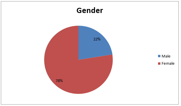
- Among group was selected, 22.8% were male where as 77.2%were female.
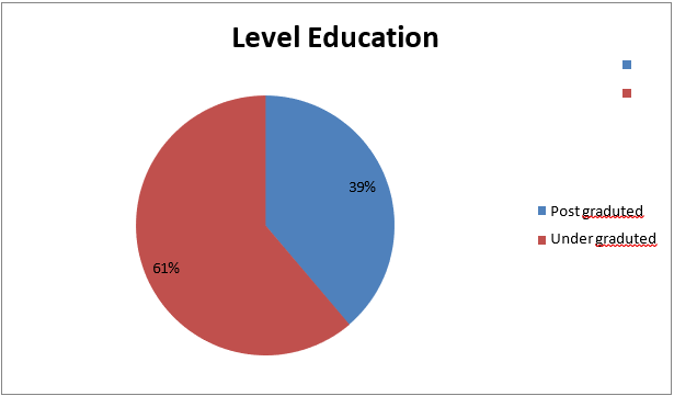
- The educational level: under graduate 62.2%, post graduate 37.8%.
- The dentist ask for X-ray for every patient about 16.5%.
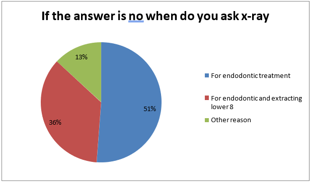
- The most dentistask for X-ray for endodontic 52%.
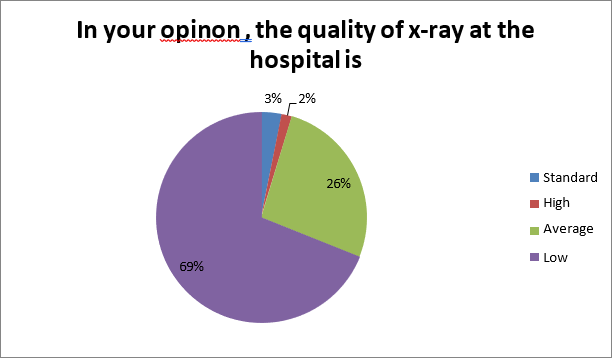
- The quality of X-ray at the hospitalis low by 69,3%.
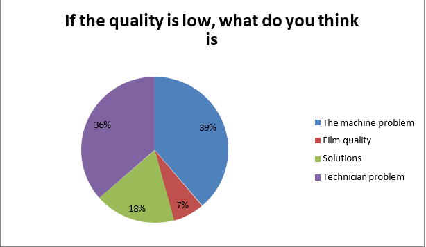
- The cause of the low quality is: the machine quality, 37.8% and the technician problem, 37%.
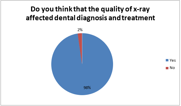
- The quality of X-ray is affects dental diagnosis and treatment about 97.6%.
Introduction
Dental X-rays (radiographs) are images of your teeth that your dentist uses to evaluate your oral health.These X-rays are used with low levels of radiationto capture images of the interior of your teeth and gums. This can help your dentist to identify problems, like cavities, tooth decay, and impacted teeth.
Dental X-rays may seem complex, but they’re actuallyvery common tools that are just as important as your teeth cleanings.
Dental X-rays are typically performed yearly. They can happen more often if your dentistis tracking the progress of a dental problem or treatment. Like brushing and flossing, getting regular dental X-rays is an integralpart of your overall oral health .
Having a good checkup can be a relief, but this doesn’t mean you shouldn’t keep getting X-rays. depending on your age, health, and insurance coverage, X-rays may be performed every one to two years. Be sure to commit to your appointments and see your dentist sooner if you experience any pain or other changes in yourmouth.
There are two main types of dental X-rays: intraoral (meaning the X-ray film is inside the mouth) and extraoral (meaning the X-ray film is outside the mouth).
Intraoral X-rays are the most common type of dental X-ray taken. These X-rays providea lot of detail and allow your dentist to find cavities,check the health of the tooth root and bone surrounding the tooth, check the status of developing teeth, and monitor the general health of your teeth and jawbone. Extraoral X-rays show teeth, but their main focus is the jaw and skull. These X-rays do not provide the detail found with intraoral X-rays and thereforeare not used for detecting cavities or for identifying problems with individual teeth. Instead, extraoral X-rays are used to look for impacted teeth, monitor growth and development of the jaws in relationto the teeth, and to identify potential problems between teeth and jaws and the temporomandibular joint (TMJ, see temporomandibular disorders for more information) or other bones of the face.
The factors that can affect the quality of radiographic images dependson:
- How the image was taken (radiographic technique).
- What image receptorwas used (filmor digital).
- How the visualimage was created.
- Chemical processing (film).
- Computer processing (digital). The high quality of dental x-rayhelp to:
- Good Diagnostic Quality.
- Consistent results.
- Lowest possibledose to patient.
- Determine sources of error and correct.
- Cost effective.
The effectsof poor radiographic technique are the same whatever type of image receptor is used. These technique errors have already been covered in detail in relation to the three main projections used in dentistry, namely periapicals, bitewings and panoramic radiographs.
Practical factorsinfluencing film-based image quality:
In practical terms, the various factors that can influence overall film-captured image quality can be dividedinto factors relatedto:
- The X-ray equipment
- The imagereceptor – film or film/screen combination
- Processing
- The patient
- The operatorand radiographic technique.
Dental care professionals need to be able to recognize the cause of the various film faults so that appropriate corrective actioncan be taken.Typical film faults.
- Overexposure owing to:
- FaultyX-ray equipment, e.g., timer
- Incorrectexposure time settingby the operator
- Overdevelopment owing to:
- Excessivetime in the developer solution
- Developersolution too hot
- Developersolution too concentrated
Fogging owing to:
- Poor storage conditions: Too warm
- Old film stock i.e. films used afterexpiry date
- Faulty cassettes allowingingress of stray light
- Faulty darkroom/processing unit:
- Allowing leakageof stray light.
- Faulty safe-light.
- Thin patienttissues.
- Underexposure owing to:
- Faulty X-ray equipment, e.g., timer.
- Incorrect exposuretime setting by the operator.
- Failure to keep timer switch depressed throughout theexposure.
- Underdevelopment owing to:
- Inadequate time in the developer solution.
- Developer solutiontoo cold.
Justification
I select the X-ray quality as a problem to be study according to many reasons:
- To evaluatethe implementation of quality assurancerequirements for digitaldental radiography in routine clinicalpracticl
- Fiercely debated
- Is the view clinically necessary
- Is there a net benefit from taking a radiograph
- Does the clinical examination warrant the x-ray
- Do the symptoms/history warrantan x-ray
- Every view must be justified and reasons recorded
Objectives:
- General objective: -
The aim of this study is to assess the quality of X ray in wad medanidantal teaching hospital and its impact on work.
- Specific objectives: -
- To gain feedback from dentists about X-ray quality.
- To set a programto improve the X-ray quality.
- To evaluatethe prevalence of the most radiograph toothat the dental hospital.
- To improveknowledge of dentistsabout the benefitsof good X-ray quality.
- To investigate the standardized criteriafor high X- ray quality.
Methodology
Study design: -
This study was a descriptive analytic cross-sectional type held at Wad madani dental teaching hospital.
Data collection and tools:
The study used a cross-sectional descriptive survey design in the form of a questionnaire of 11 questions about: (demographic data, evaluation of dental x-ray in hospital, quality of dental radiograph, related b/w low quality of x-ray and misdiagnosis, prevelance of most radiograph tooth).
Site:
Various places in Wad madani dental teaching hospital and randomly selected.
Target Population:
Dentist in Wad madani dental teaching hospital.
Sampling and sample frame:
Simple random sampling was used to select my sample.
Selection Criteria:
Inclusion criteria: Dentist in wad medani dental hospital between under graduate and post graduate who agree to participate in the study.
Exclusion criteria:
No exclusion in may sample
Ethical Considerations:
All study participants gave informed consent prior to participating in the study which was conducted on an anonymous basis.
Data Analysis:
Data was been analysis using manual method.
Literature Review
- "1" Dental practitioners in the United States are exposing more than 400 million dental radiographs per year. Major nationalstudies have indicatedthat improvements in radiographic qualityis needed. Qualityassurance (QA) programsused in medical radiology are designed to produce radiographs that are of high quality, use the least amount of radiation, and are produced at minimum cost. Preliminary recommendations are presentedfrom the QualityAssurance Committee of the AmericanAcademy of Dental Radiology as an outlinefor establishing preventive maintenance of x-ray systems and a preliminary method for determining appropriate QA monitoring levels for dentistry. Included are recommendations for three stages of dental radiology QA. These are preliminary ratings and are expected to change as more dental QA information becomes available. “The American Academy of Dental Radiology Quality Assurance Committee 15 May 2005”.
- "2" An adequate quality radiograph is one, which provides the required diagnostic information. However, the quality of radiograph depends upon several contributory factors. Where the practioners is in any doubt about the reasons for poor radiographic quality It is helpful to systematically target the problem areas. This is achieved by carrying out a film reject analysis. “Shams Ul Nisa (Author)2016”
- "3" An organised effort by the staff operating a facility to ensure that the diagnostic images produced by the facility are of a sufficiently high quality so that they consistently provide diagnostic information at the lowest possible cost and with the least possibleexposure of the patient to radiation. “WHO 2005”.
- "4" Radiationdosimetry for determining the optimum image quality with the lowest radiation exposure to the patient was carried out. The best image quality with the lowest exposure dose was assessedfor conventional intraoralX-ray film (Kodak type E) and the digital processing sensor (RVG 5200). Radiation surveylevel was done during this study for safety and protection Purposes. “King Abdul AzizUniversity (KAU) Dental Hospital”.
- "5" Quality assurance programmes for dental radiography are needed to ensure that images are consistently of a high standard. All aspects of the imaging process must be monitored to reduce the number of repeat radiographs needed, and to ensure radiography is carried out efficiently.” Jane Luker 2014”.
- "6"The purposeof Quality Assurance(QA) in dental radiology is to ensure consistently adequatediagnostic information, while radiation doses are controlled to be as low as reasonably achievable. “1996. European Guidelines on Quality criteria”.
Results of data analysis


Results
-The study sample consistof post graduate 38%, under graduate 61%, 22.5% were Males and 77.5% were Females.
-Most dentistare not ask for x-ray for every patient (82.9%).
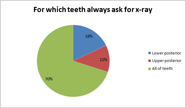
-Most ask for x-ray in endodontic reason (51.2%) to the all teeth (69%).
-The qualityof x-ray is low (69%).
-In this sample, (97.7%)low x-ray quality effect of diagnosis and treatment.
-In this sample the main cause of low quality is machineproblems (38.8%).
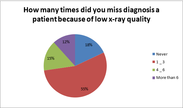
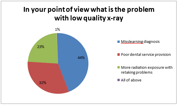
-In this sample the main problemwith low qualityof x-ray is misleading diagnosis (44.2%).
Discussion
-This study revealed that assesment of x-ray qualitywad medani dental teaching hospital assest the quality in study area was 69% low and 26.4% average which is higher than the most previous study.
-This difference may be attributed to the difference in quality of x-ray, methodology, misdiagnosis effects as well as failure RCT and difficult extracting lower 8.
-As a result of all these variables, film faults and alterations in image quality are inevitable. However, since the diagnostic yield from radiography is related directlyto the quality of the image, regular checks and monitoring of these variablesare essential to achieve and improve the quality of radiographs. It is these checks which form the basis of quality assurance programmers.
Conclusion
In wad medani dental teaching hospital the quality of x-ray is low (69%), the main causes is machine quality problem (38.8%) and technician problem (36.4%).
Recommendation
-Improve the machine qualityand using with profession technician.
-Improve the image and soluation quality.
-The technician must respect and adhere to the workinghours.
-There is an increased demand to start educational programsfor dental student and dentist and teachers to increased their dentalknowledge and management about radiograph machine.
-Select the high quality of solutions and good type of film, using the criteria given for film quality.
-As a minimum target,no greater than 20% of radiographs shouldbe of unacceptable quality.
-As soon as possible,insert the digitalx-ray machine.
References
- E.T. Parks, G.F. Williamson. (2002). “Digital radiography: an overview.” J Contemp Dent Pract 3:23–39.
- P.F. Van Der Stelt. (2005). “Filmless imaging. The uses of digital radiography in dental practice.” JADA.
- E.T. Parks. “Digital radiographic imaging. Is the dental practice ready?” JADA 139:447–481 (2008).
- G.L. Conover, C.F. Hildebolt, D. (1995). Anthony. “Objective and subjective evaluations of Kodak Ekta-speed Plus dental x-ray film.” Oral Surg Oral, Med Oral Pathol Oral Radiol Endod 79:246–250.
- B. Svenson, U. Welander, X.Q. Shi, H. Stamatakis, G. (1997). Tronje. “A sensitometric comparison of four dental X-ray films and their diagnostic accuracy.” Dentomaxillofac Radiol 26(4): 230–235.
- E.M. Tjelmeland, W.S. Moore, C.B. Hermesch, D.J. Buikema. (1998). “A perceptibility curve comparison of Ultra-speed and Ektaspeed Plus films.” Oral Surg Oral Med Oral Pathol Oral Radiol Endod 85: 488–485
- K. Syriopoulos, X.L. Velders, G.C. Sanderink, P.F. (2001). Van Der Stelt. “Sensitometric and clinical evalu-ation of a new F-speed dental X-ray film.” Dentomaxillofac Radiol 30:40–44.
- D.I. Bernstein, S.J. Clark, J.P. Scheetz, A.G. Farman, B. Rosenson. (2003). “Perceived quality of radio-graphic images after rapid processing of D- and F-speed direct-exposure intraoral x- ray films”. Oral Surg Oral Med Oral Pathol Oral Radiol 96:486–491.
- National Council on Radiation Protection and Measurements, NCRP report 145: Radiation Protec-tion in Dentistry. National Council on Radiation Protection and Measurements, Bethesda, MD,2003.
- American Dental Association Council on Scientific Affairs. “The use of dental radiographs: Updateand recommendations American Dental Association Council on Scientific Affairs.” JADA 137
- National Council on Radiation Protection and Measurements. NCRP Report 172: Reference Levels and Achievable Doses in Medical and Dental Imaging: Recommendations for the United States.
- National Council on Radiation Protection and Measurements, Bethesda, MD, 2012.


