Archive : Article / Volume 2, Issue 1
- Case Report | DOI:
- https://doi.org/10.58489/2836-5127/005
Using CT scan to Detect Radiolucent Foreign Body (Glass): An Experimental Study
Radiology Specialist, Department of Radiology, Al-Namas General Hospital, Ministry of Health, Al-Namas City, Saudi Arabia
Abdulwahab F. Alahmari
Abdulwahab F. Alahmari, (2023). Using CT scan to Detect Radiolucent Foreign Body (Glass): An Experimental Study. Journal of Radiology Research and Diagnostic Imaging. 2(1). DOI: 10.58489/2836-5127/005.
© 2023, Abdulwahab F. Alahmari, this is an open access article distributed under the Creative Commons Attribution License, which permits unrestricted use, distribution, and reproduction in any medium, provided the original work is properly cited.
- Received Date: 13-01-2023
- Accepted Date: 22-01-2023
- Published Date: 28-01-2023
Computed Tomography; Radiolucent Foreign Body; Color Table; Detection; Translucent.
Abstract
Usually on X-ray of the foot or the hand to detect a translucent foreign body (glass) do not work most the time. As well, ultrasound was used to detect some glass types before. The aim of this paper is to use CT scan and the color lookup table to detect the glass near a phantom.
Introduction
There are 66 types of glasses [1] and maybe more. Some of these glasses are undetectable on an X-ray even though many claims all glasses are detectable on X-ray [1]. Other disagree, and they claim that only 10% of the radiographs give positive feedback, but it can detect 85% of the radiopaque glass [2]. There are many factors that affect the detectability of the glass: 1-Type of glass, 2-Density of the glass, 3-Location of the glass, 4-Size of the glass, 5-The percentage of lead and silica in the glass. Even thou; other claim that led and silica percentages in glass are not important factors [3, 4]. Other papers claim that only glass that is bigger than 2 mm will be detected on radiographs [5,6,7]. In another paper, the authors claim all glass is radiopaque [3]. Another author claim that all glass is detectable on CT scan [8]. The density of the glass varies between 500-1900 HU with average of 1200 [8]. Any dense fragment that is 0.01 is detectable on CT scan [7]. Ultrasound can detect glass, but it will cause reverberation artifact that appears as a posterior acoustic shadowing [7]. On MRI glass is seen on all sequences, but artifact is present too, so MRI should not be considered [8].
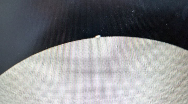
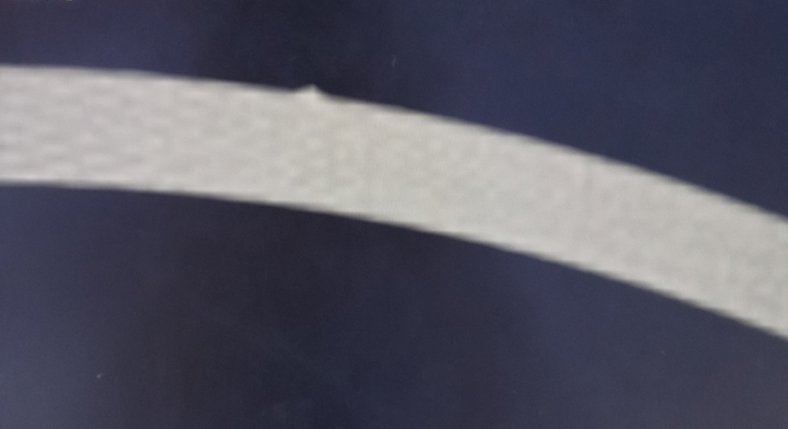
The Experiment and The Result
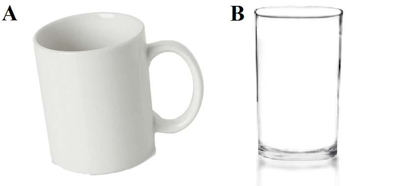
In this paper, 2 pieces of glass were taken one from a white glass cup and one from a transparent glass. The pieces were broken as small as possible. Both types of glass were put on a phantom and scanned by the CT scanner. The colors lookup table were used with both to see if it will make them distinct from the phantom. The 1st glass size is 0.09 mm and the 2nd glass size is 0.05 mm. the first glass was radiopaque and easily to be identified. The second glass was radiolucent and its texture not distinct from the phantom texture. To conclude the experiment, the glass type and size are very important to find the foreign body in the patient. As well, the colors lookup table did not provide a great help. The size 0.05 mm were seen on the CT scan, but the low density of the glass made it impossible to differentiate the glass texture from the phantom texture. X-ray and CT scan can’t show all types of glass. Therefore, ultrasound might provide a solution. The amount of lead and silica used in the glass increase as the density/Hounsfield unit increase and appeared as a radiopaque in the scan.
Discussion
Accordingto many papers, all glasses are visible on CT scan [1,3,8], which proven in this paper to be wrong. A simple test using a transparent glass was placed on a phantom and scanned by a CT scanner did not show different density for the glass from the phantom. Ultrasound is superior to CT scan in imaging glass which show acoustic shadowing [4], meanwhile, CT scan can’t show some types of glasses as found in this paper. The glass depth in the body will determine the ability of ultrasound ability to locate the glass.
Conclusion
Ultrasound is superior to CT scanner in imaging foreign body (i.e., glass) and a comparison study must be done. Some authors claim that CT scan can show any type of glass which is wrong statement and exaggeration. Color lookup table did not help in detecting the glass.
References
- Tandberg, D. (1982). Glass in the hand and foot: Will an x-ray film show it? JAMA, 248(15), 1872-1874.
- Skinner, D., Skinner, D. V., Swain, A., Robertson, C., & Peyton, J. R. (Eds.). (1997). Cambridge textbook of accident and emergency medicine. Cambridge University Press.
- Klein, K. A., & Hobbs, B. B. (1995). Radiopacity of glass: does the lead content matter? CMAJ: Canadian Medical Association Journal, 153(9), 1224.
- Aras, M. H., Miloglu, O., Barutcugil, C., Kantarci, M., Ozcan, E., & Harorli, A. (2010). Comparison of the sensitivity for detecting foreign bodies among conventional plain radiography, computed tomography and ultrasonography. Dentomaxillofacial Radiology, 39(2), 72-78.
- Hunter, T. B., & Taljanovic, M. S. (2003). Foreign bodies. Radiographics, 23(3), 731-757.
- Halaas, G. W. (2007). Management of foreign bodies in the skin. American family physician, 76(5), 683-688.
- Carneiro, B. C., Cruz, I. A., Chemin, R. N., Rizzetto, T. A., Guimarães, J. B., Silva, F. D., ... & Nico, M. A. (2020). Multimodality imaging of foreign bodies: new insights into old challenges. Radiographics, 40(7), 1965-1986.
- Ingraham, C. R., Mannelli, L., Robinson, J. D., & Linnau, K. F. (2015). Radiology of foreign bodies: how do we image them? Emergency radiology, 22(4), 425-430.


