Archive : Article / Volume 1, Issue 1
Case Report | DOI: https://doi.org/10.58489/2836-2217/002
The Role of The Hippocampus in Conditionally Reflective Activity and The Formation of The Functional State of The Crust of The Large Hemisphere
1.Olena Lutsenko Assistant Lecturer of the Department of Theory and Methods of Teaching of Natural Sciences, Hlukhiv National Pedagogical University of Alexander Dovzhenko, Hlukhiv, Ukraine.
2.Svitlana Furtatova Phd, Associate Professor of the Department Anatomy of Physiology and Physical Rehabilitation of Bohdan Khmelnytsky Cherkasy National University, Cherkasy, Ukraine.
Correspondng Author: Olena Lutsenko
Citation: Olena. L, Svitlana. F, (2022). The Role of the Hippocampus in Conditionally Reflective Activity and the Formation of the Functional State of the Crust of the Large Hemisphere. Journal of Clinical Case Reports and Trails. 1(1). DOI: 10.58489/2836-2217/002
Copyright: © 2022 Olena Lutsenko, this is an open access article distributed under the Creative Commons Attribution License, which permits unrestricted use, distribution, and reproduction in any medium, provided the original work is properly cited.
Received Date: 2022-07-08, Received Date: 2022-07-08, Published Date: 2022-08-18
Abstract Keywords: ventral hippocampus, dorsal hippocampus, retarded inhibition, conditional reflex activity, functional state of the cerebral cortex, electrical stimulation.
Abstract
The studies were carried out on dogs, which were stereotactically implanted with depth electrodes in the ventral and dorsal parts of the hippocampus. We studied the effect of stimulation of these structures with an electric current of high (60 pulses/sec) and low (4 pulses/sec) frequencies on conditional reflex activity and the formation of the functional state of the cerebral cortex. It was revealed that stimulation of different structures of the hippocampus has a different effect on the tone of cortex cells. The nature of this influence depends on the frequency of the electric current, which was used to stimulate.
Formulation of the problem. Analysis of recent research and publications. There are a number of scientific papers devoted to the study of the role of the hippocampus in the regulation of various body functions (Giuliana, Caronia, Jennifer, Wilcoxon, et all., 2010; Purvis, EM, O'Donnelly, JC et all, 2020). Some authors believe that the hippocampus is a poly-functional structure: the center of visceral functions and sense of smell, memory, and learning, the regulator of emotions and motivations, it is used for orientation reflexes and attention, sensory analysis (Chaychenko, G.M., 1984; Wiskott L, Rasch MJ, Kempermann G. Wiskott L, Rasch MJ, Kempermann G. et all. 2006). But today there is not enough reliable data on the role of this structure in conditioned reflex activity under conditions of electrical stimulation of various structures of the hippocampus. It was found that when the hippocampus is stimulated by a weak electric current during the formation of conditioned reflexes, the fixation time of the reflex is significantly reduced, ie the processes of memorization and learning are accelerated (Gray, J.A., 1983; Diana, X Yu, Maria, C Marchetto, Fred, H Gage, 2014; Nader, K, Krysiak A, Beroun, A, et all., 2019; Whitfield, JF, Chakravarthy, BR., 2009). The mechanism of temporal connections has been shown to play an important role in regulating cortical tone. If the question of the formation of the functional state of the cortex of the large hemispheres during the formation of conditioned reflexes is more or less studied, then the question of the dependence of this phenomenon on the properties of the inhibitory process is insufficiently studied (William, D Jones, Sarah, M Guadiana, Elizabeth, A Grove, 2019). In addition, the role of various subcortical structures and, in particular, the hippocampus in the regulation of the tone of the cortex of the large hemispheres has not been clarified to date.
The purpose to establish features of the formation of a functional condition of the bark of large hemispheres in the conditions of electric stimulation of various structures of a hippocampus.
Research methods
Implantation of bipolar irritating electrodes was performed by the stereotactic method. Deep electrodes in this way we're introduced to dogs in the ventral and dorsal parts of the hippocampus.
Before and after the operation, the background indicators of conditioned reflexes, their latent periods, and the inactive phase of the delayed reflex, as well as the threshold value of electrical stimulation were determined in animals. Then, studies were conducted to study the stimulation of the hippocampus by the electric current on the conditioned reflex activity of dogs and their behavior.
In our experiments, a rectangular current with an amplitude of 3 V, a frequency of 4 pulses / s and 60 pulses / s was used. The duration of electrical stimulation was 10s. In the same animal, the stimulation of the hippocampal structures with a single current frequency was performed 12 times, namely, until a clear dependence of the functional state of the cortex of the large hemispheres on the parameters of the irritating current.
Research results and their discussion
Before analyzing the materials obtained as a result of stimulation of the hippocampus by an electric current of different frequencies, it is necessary to indicate changes in the behavior of animals during the experiments. After stimulation of the dorsal and ventral hippocampus with a current of 60 pulses/sec in most cases, the animals were too excited, very anxious, which was manifested in trembling limbs, sharp movements of the head, pronounced orientation reflex, whining, frequent yawning dogs. After stimulation of the above structures of the hippocampus with a current frequency of 4 imp / s, these deviations in behavior were not detected. Animals were even calmer on days of stimulation than on days without stimulation.
Our indicators showed (Fig. 1) that stimulation of both dorsal (A) and ventral (B) hippocampus with a high-frequency current of 60 imp / helped to increase the value of all conditioned reflexes to different stimuli compared to their background value, but after stimulation of dorsal hippocampus, it was higher than after stimulation of the ventral hippocampus with a current of this frequency. At the same time, stimulation of the hippocampus with a low-frequency current of 4 pulses / sec reduced the magnitude of conditioned reflexes to sound (except for the call reflex) and light stimuli.
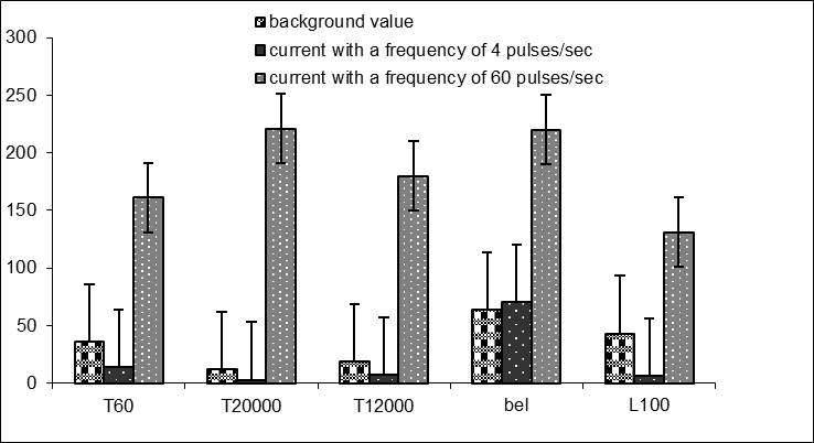
Horizontally - a kind of conditioned reflex
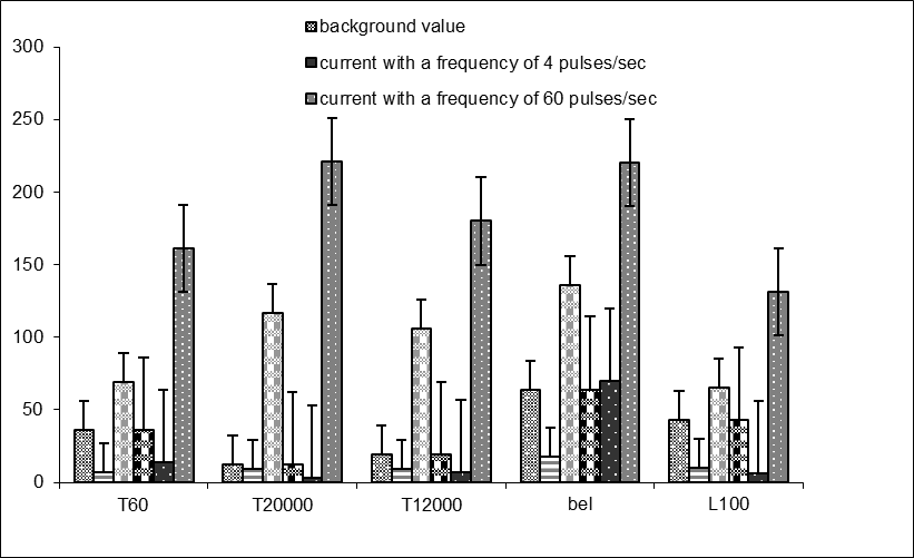
The fact that the hippocampus affects the functional state of cortical cells can be judged by the change in the magnitude of the latent periods of conditioned reflexes (Fig. 2).
In our experiments under conditions of high-frequency hippocampal stimulation, the excitability of the cerebral cortex was significantly higher than after low-frequency current stimulation, and after dorsal hippocampal stimulation the excitability of cortical cells of auditory and visual analyzers was higher than in ventral hippocampal stimulation.

A
B
Vertically - the value of latent periods in seconds
Horizontally - a kind of conditioned stimulus
*Stimulation of dorsal (A) and ventral (B) hippocampus
It should be noted that irritation of the ventral hippocampus with a current of 4 imp/sec contributed to a smaller decrease in the excitability of cortical cells than the irritation of the dorsal.
Stimulation of various hippocampal structures affected the delayed conditioned reflex. Thus, the use of low-frequency current of 4 pulses/sec led to an increase in the braking process. The magnitude of the inactive phase of the delayed conditioned reflex increased, but in conditions of dorsal hippocampal irritation more than in conditions of ventral irritation (inactive phase increased from 25 s background values to 33 s and 25 s, respectively), after stimulation of the above hippocampal sections with high frequency 60 imp/sec inactive phase in both cases decreased and this decrease after stimulation of the dorsal hippocampus was greater than after stimulation of the ventral.
The magnitude of the delayed conditioned reflex after the application of a high-frequency current of 60 imp/sec increased both after stimulation of the dorsal and ventral hippocampus but in the first case much more than in the second. We observed an increase in the value of the delayed reflex when using a low-frequency current of 4 pulses / s only when the ventral hippocampus is irritated. Under conditions of irritation of the dorsal hippocampus by a current of this frequency, there was a decrease in the magnitude of the delayed reflex.
Under the influence of hippocampal stimulation, the functional state of the cells changed not only of the signal analyzer but also of the state of the cells responsible for the perception of the unconditioned stimulus.
In most cases, the threshold value of the unconditioned reflex after hippocampal stimulation increased, and after low-frequency current stimulation more than in high-frequency current stimulation, and under dorsal hippocampal stimulation, this increase was more significant than under ventral stimulation. After stimulation of the latter with a high frequency current of 60 pulses / sec, the threshold value of the unconditioned reflex remained at the same level as before stimulation (0.01 mA).
Thus, our studies suggest that changes in the functional state of cortical cells, which occurred during the formation of conditioned reflexes in dogs, significantly depended on the ascending effects of such a subcortical structure as the hippocampus. As the fixation of conditioned reflexes and especially under the influence of internal inhibition, the optimal level of the functional state of the central nervous system is maintained and reproduced by the conditioned reflex mechanism formed in the cortex of the large hemispheres.
Conclusions
1. Stimulation of the dorsal hippocampus with a high frequency current of 60 imp / s contributed to a greater increase in the value of positive conditioned reflexes and reduce their latency periods than the stimulation of the ventral. Stimulation of both parts of the hippocampus with a low-frequency current of 4 pulses / s did not significantly affect the magnitude of conditioned reflexes produced by sound and light stimuli.
2. Irritation of the dorsal hippocampus by low-frequency current led to the suppression of the delayed reflex, while irritation of the ventral - to increase.
3. The inhibitory phase of the delayed conditioned reflex with the application of low-frequency current increased in comparison with the background value under conditions of stimulation of both structures of the hippocampus to a greater extent than the ventral. Stimulation of the dorsal hippocampus with high-frequency current caused a weakening of the inhibitory phase of the delayed reflex more significantly than ventral irritation.
4. The threshold value of unconditional stimuli was significantly increased by the stimulation of various hippocampal structures with low-frequency current (4 pulses / s), while the use of high frequency current (60 pulses / s) had little effect on the functional state of the center of unconditional reflex.
References
- Giuliana, Caronia, Jennifer, Wilcoxon, Polina, Feldman, Elizabeth, A Grove (2010) Bone morphogenetic protein signaling in the developing telencephalon controls formation of the hippocampal dentate gyrus and modifies fear-related behavior J Neurosci. May 5;30(18):6291-301. doi: 10.1523/JNEUROSCI.0550-10.2010.
- Diana, X Yu, Maria, C Marchetto, Fred, H Gage (2014) How to make a hippocampal dentate gyrus granule neuron Development. Jun;141(12):2366-75. doi: 10.1242/dev.096776.
- Christiana, G Martin, Hyungsuk, Kim, Sijung, Yun, Whitney, Livingston, Joseph, Fetta, Vincent, Mysliwiec, Tristin, Baxter, Jessica, M Gill (2017). Circulating miRNA associated with posttraumatic stress disorder in a cohort of military combat veterans Psychiatry Res. Р. 251:261-265. doi: 10.1016/j.psychres.2017.01.081.
- William, D Jones, Sarah, M Guadiana, Elizabeth, A Grove (2019). A model of neocortical area patterning in the lissencephalic mouse may hold for larger gyrencephalic brains J Comp Neurol. Vol. 15;527(9): 1461-1477. doi: 10.1002/cne.24643.
- Whitfield, JF, Chakravarthy, BR. (2009) The neuronal primary cilium: driver of neurogenesis and memory formation in the hippocampal dentate gyrus? Cell Signal. Vol 21(9):1351-5. doi: 10.1016/j.cellsig.2009.02.013.
- Purvis, EM, O'Donnelly, JC, Chen, HI, Cullen, DK (2020) Tissue Engineering and Biomaterial Strategies to Elicit Endogenous Neuronal Replacement in the Brain. Front Neurol. 2020 Apr 28; 11:344. doi: 10.3389/fneur.2020.00344
- Nader, K, Krysiak A, Beroun, A, Pekala M, Szymanska, M, Kuzniewska, B, Radwanska, K, Kaczmarek, L, Kalita, K. (2019) Loss of serum response factor in mature neurons in the dentate gyrus alters the morphology of dendritic spines and hippocampus-dependent behavioral tasks. Brain Struct Funct. 2019 Nov; 224(8):2691-2701. doi: 10.1007/s00429-019-01925-6.
- Wiskott L, Rasch MJ, Kempermann G. (2006) A functional hypothesis for adult hippocampal neurogenesis: avoidance of catastrophic interference in the dentate gyrus. Hippocampus. Vol. 16(3):329-43. doi: 10.1002/hipo.20167.
- Chaychenko, G.M. Functional role of dorsal and ventral hippocampus in olfactory behavior of rats. Journal of Higher Nervous Activity. 1984. Vol. 34, №6. P. 1109-1115 (in Ukraine)
- Gray, J.A. Comparison between the behavioural effecrs of septal and hippocampal lesions: a review. Neurosci. And Biobehav. Rew. McNaughton N, 1983, v.7, n2, p.119


