Current Issue : Article / Volume 3, Issue 1
- CLINICAL IMAGES | DOI:
- https://doi.org/10.58489/2836-5127/020
Coup-Contrecoup Brain Injury: Clinical Images
- Radiology Specialist, Radiology Department, Al-Namas General Hospital, Ministry of Health, Al-Namas City, Saudi Arabia.
Abdulwahab F. Alahmari
Abdulwahab F. Alahmari. (2024). Coup-Contrecoup Brain Injury: Clinical Images. Radiology Research and Diagnostic Imaging. 3(1); DOI: 10.58489/2836-5127/020
© 2024 Abdulwahab F. Alahmari, this is an open-access article distributed under the Creative Commons Attribution License, which permits unrestricted use, distribution, and reproduction in any medium, provided the original work is properly cited.
- Received Date: 23-05-2024
- Accepted Date: 01-06-2024
- Published Date: 04-06-2024
Abstract
Introduction
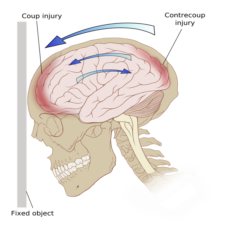
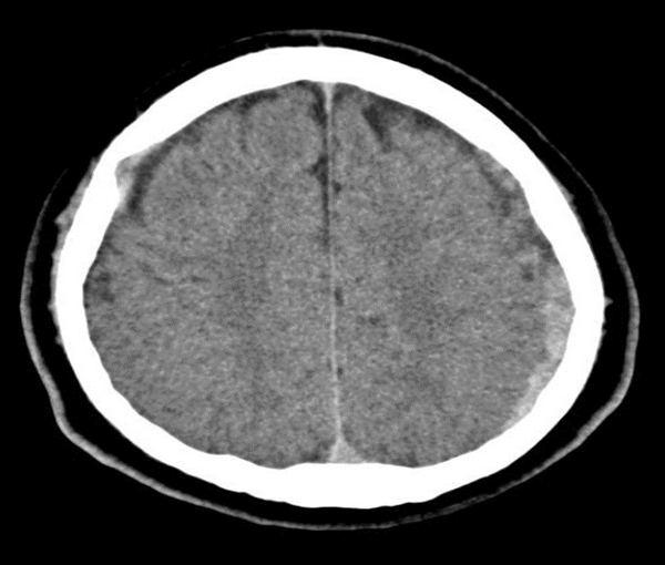
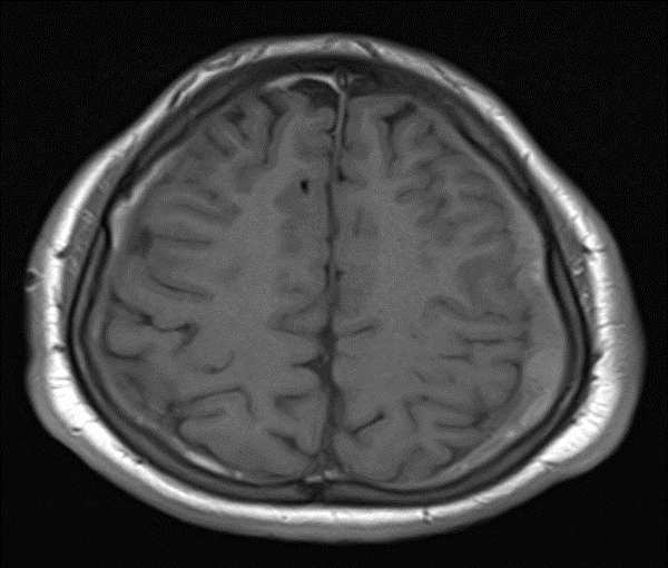
Coup-Contrecoup Brain Injury is when a part of the brain is injured and the opposite part of the brain deaccelerates and hits the inner table of the skull see (Fig. 1). This is a case of a Coup-Contrecoup Brain Injury in a 17-year-old male patient. The trauma targeted the right frontal lobe and the left parietal lobe was affected by the trauma see (Figs. 2 and 3). The patient was unstable and shifted to a tertiary hospital. Coup-contrecoup brain injuries were studied in two Indian studies [1, 2], one for 74 patients over the course of one year. The other paper studied 298 patients over a 2-year period. The first study showed that contrecoup contusion leads to a poor prognosis [1]. The second study showed that there is a difference in mortality between coup-contrecoup injuries and contrecoup injuries [2].
References
- Bhateja, A., Shukla, D., Devi, B. I., & Kolluri, V. S. (2009). Coup and contrecoup head injuries: Predictors of outcome. Indian Journal of Neurotrauma, 6(02), 115-118.
- Banga MS, Sandeep BV, Roy K, Saha SK, Dixit S, Ghosh P. Contrecoup head injury. Indian Journal of Neurosurgery. 2017 Aug;6(02):103-6.


